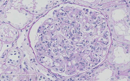
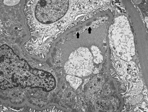
1,838
Views
5
CrossRef citations to date
0
Altmetric
Case Report

Human Parvovirus B19-Induced Acute Glomerulonephritis: A Case Report

, , , , , , , & show all
Pages 159-162 | Received 09 Jul 2012, Accepted 27 Sep 2012, Published online: 01 Nov 2012

Figures & data

Figure 1.  Light microscopy findings. Light microscopy shows a glomerular lesion that was enlarged and hypercellular (PAS staining, original magnification ×200).

Figure 1.  Light microscopy findings. Light microscopy shows a glomerular lesion that was enlarged and hypercellular (PAS staining, original magnification ×200).

Figure 2.  Electron microscopy findings. The electron-dense deposits (arrows) were detected in the subendothelial area and mesangial area but not in the subepithelial area.

Figure 2.  Electron microscopy findings. The electron-dense deposits (arrows) were detected in the subendothelial area and mesangial area but not in the subepithelial area.

Table 1.  The characteristics of patients with endocapillary proliferative glomerulonephritis induced by HPVB19 infection.

Reprints and Corporate Permissions

Please note: Selecting permissions does not provide access to the full text of the article, please see our help page How do I view content?

To request a reprint or corporate permissions for this article, please click on the relevant link below:

Academic Permissions

Please note: Selecting permissions does not provide access to the full text of the article, please see our help page How do I view content?

Obtain permissions instantly via Rightslink by clicking on the button below:

If you are unable to obtain permissions via Rightslink, please complete and submit this Permissions form. For more information, please visit our Permissions help page.