
Figures & data
Figure 1. Flow cytometric analysis of lymphocyte populations: (A) Forward and side scatter histogram was used to define the lymphocyte population (R1). (B) The expression of B and T cell markers were assessed in the lymphocyte population as CD4+ and CD8+ compared with the negative isotype control (not shown). (C) CD19+ cells were gated. (D, E) The expression of CD5 and CD27 in B cells was detected.
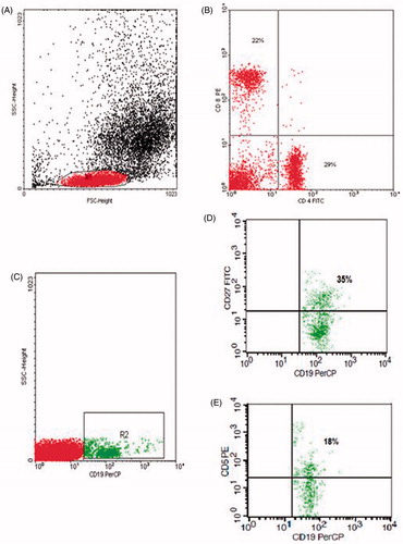
Figure 2. Flow cytometric detection of apoptosis of B and T lymphocytes: (A) Forward and side scatter histogram was used to define the lymphocyte population (R1). (B and C) CD19+ B cells and CD3+ T cells were gated. (D) Annexin V expression on lymphocyte populations. The positivity was defined as fluorescence (grey (green) histogram) higher than that of the isotype control (black (blue) histogram).
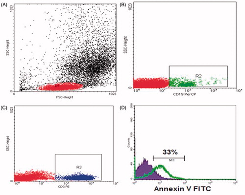
Table 1. Baseline clinical and biochemical characteristics of patients with ESRD and controls.
Table 2. Leukocyte count and lymphocyte subsets in patients with ESRD and controls.
Table 3. Apoptosis of B lymphocytes and T lymphocytes in patients with ESRD and controls.