Figures & data
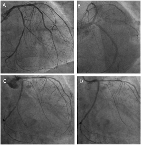
Figure 1. Panel A: coronary angiogram showing mild stenosis of mid-LAD and severe stenosis at proximal segment of dominant LCX; Panel B: angiographic image after bare metal stent implantation on proximal LCX; Panel C: coronary angiogram showing severe stenosis at the ostium of LM, multiple stenosis of middle LAD, first diagonal branches, and in-stent restenosis of LCX involving obtuse marginal branch. Panel D: angiogram showing occlusion of both by-pass grafts; Panel E: native coronary artery angiogram showing severe stenosis of ostial LM, moderate stenosis of mid-LAD, with diffuse disease of diagonal and septal branches; moderate in-stent restenosis on proximal LCX and severe; Panel F: angiographic image after bare metal stent implantation on LM. LAD, left anterior descending artery; LCX, left circumflex artery; LM, left main; NSTEMI, non-ST elevation myocardial infarction; PL, posterolateral branch.
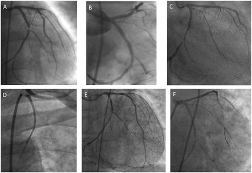
Figure 2. Panel A: coronary angiogram showing focal in-stent restenosis of LM, tight stenosis of ostial LCX, and a long lesion in distal LCX beyond the distal edge of the previous implanted stent. Panel B: angiographic image after multiple bare metal stent implantations on proximal and distal LCX, and in-stent re-PTCA of LM. Panel C: coronary angiogram showing severe and diffuse in-stent restenosis of LCX involving the ostium of OM branch; Panel D: angiographic image after re-PTCA of LCX.