
Figures & data
Table 1. Laboratory data at presentation.
Figure 1. Renal biopsy findings. (A) Glomerular basement membranes appeared uniformly thickened without spikes (stained by periodic methenamine silver, original magnification ×400). (B) Tiny fuchsinophilic grains deposits along the outer aspect of the glomerular basement membranes (stained with Masson’s trichrome, original magnification ×400). (C) Immunoglobulin G subclasses IgG1 deposited along the glomerular capillary wall (stained immunohistochemically, original magnification ×400). (D) Immunoglobulin G subclasses IgG3 deposited along the glomerular capillary wall (stained immunohistochemically, original magnification ×400). (E) Effacement and fusion of epithelial cell foot processes (electron microscopy; original magnification ×8000). (F) Presence of subepithelial immune complex deposits (electron microscopy; original magnification ×10,000).
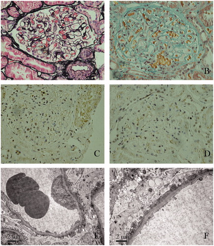
Figure 2. Clinical course after hospitalization. Time course of clinical indices after admission (A) serum total protein; (B) serum albumin; (C) proteinuria.
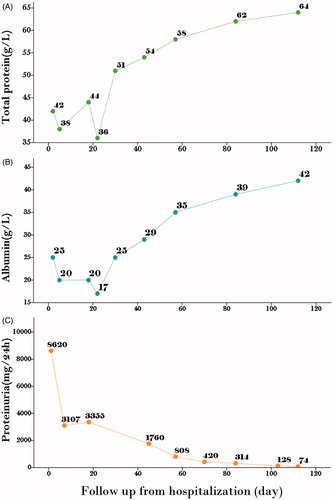
Table 2. Tiopronin-induced nephrotic syndrome cases reported in the literature.