
Figures & data
Figure 1. Intrarenal localization and roles of the CaSR. Notes: Cellular polarity of the CaSR is apical [in the proximal tubule and outer/inner medullary collecting duct (OMCD/IMCD)] and basolateral [in the thick ascending limb (TAL) and, occasionally, in the cortical collecting duct (CCD)]. Species differences exist in the distal convoluted tubule (DCT)/connecting segment (CNT), where receptor expression can be detected apically and/or basolaterally/intracellularly. PCT/PST, proximal convoluted/straight tubule; tDL/tAL, thin discending/ascending limb; MTAL/CTAL, medullary/cortical thick ascending limb; JGA, juxtaglomerular apparatus; TRPV5, transient receptor potential vanilloid 5; PMCA, plasma membrane Ca2_-ATPase; VitD, vitamin D; VDR, vitamin D receptor; NKCC2, Na_-K_-2Cl_ cotransporter 2; ROMK, renal outer medullary potassium K_ channel; PTH, parathyroid hormone; AQP2, aquaporin 2.
![Figure 1. Intrarenal localization and roles of the CaSR. Notes: Cellular polarity of the CaSR is apical [in the proximal tubule and outer/inner medullary collecting duct (OMCD/IMCD)] and basolateral [in the thick ascending limb (TAL) and, occasionally, in the cortical collecting duct (CCD)]. Species differences exist in the distal convoluted tubule (DCT)/connecting segment (CNT), where receptor expression can be detected apically and/or basolaterally/intracellularly. PCT/PST, proximal convoluted/straight tubule; tDL/tAL, thin discending/ascending limb; MTAL/CTAL, medullary/cortical thick ascending limb; JGA, juxtaglomerular apparatus; TRPV5, transient receptor potential vanilloid 5; PMCA, plasma membrane Ca2_-ATPase; VitD, vitamin D; VDR, vitamin D receptor; NKCC2, Na_-K_-2Cl_ cotransporter 2; ROMK, renal outer medullary potassium K_ channel; PTH, parathyroid hormone; AQP2, aquaporin 2.](/cms/asset/e5d12589-92e4-4bf8-973d-a66e2a672b6a/irnf_a_937673_f0001_b.jpg)
Figure 2. CaSR activities in a cell model from the thick ascending limb (TAL). Notes: CaSR decreases paracellular calcium reabsorption by inhibition of renal outer medullary potassium channel (ROMK)-mediated potassium recycling to the lumen that supports the luminal positive electric gradient. This gradient is the driving force for the passive paracellular calcium reabsorption in the TAL. AA, arachidonic acid; 20-HETE, 20-hydroxyeicosatetraenoic acid; PKA, protein kinase A; PLA2, phospholipase A2.
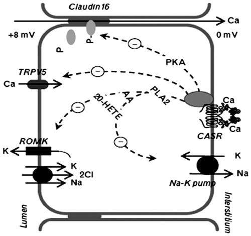
Figure 3. CaSR in distal convoluted tubule. CaSR reduces calcium pump activity (PMCA) and calcium active reabsorption.
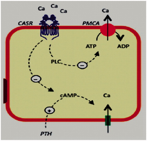
Table 1. The associated studies regarding calcium sensing receptor gene polymorphisms in patients with nephrolithiasis.