Figures & data
Table 1. The findings of laboratory tests performed on admission.
Figure 1. The light microscopic findings. (A) While globally sclerosed glomeruli and collapsed glomeruli were observed, the other glomeruli showed an almost normal appearance (Masson trichrome stain, original magnification ×40). (B) This glomerulus looks normal. However, the vascular smooth muscle cells are disorganized and enlarged (PAM-HE stain, original magnification ×200). (C) This glomerulus also looks normal, while the myocytes of the afferent arteriole are enlarged and their endothelium has slight hyalinosis (PAM-HE stain, original magnification ×200). (D) Granular swollen epithelial cells are indicated by arrows (Masson trichrome stain, original magnification ×400).
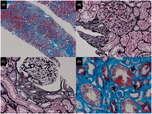
Figure 2. The electron microscopic findings. (A) The foot processes of podocytes are effaced (asterisks). (B) The granular swollen epithelial cells have increased numbers of mitochondria, a portion of which are enlarged, and some of which have cristae that have lost their normal structure.
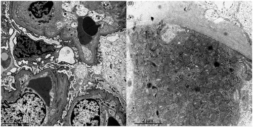
Figure 3. The results of the direct sequencing of mtDNA. A homoplasmic 7501 T > A variant in the tRNASer(UCN) was detected in the patient’s blood sample.
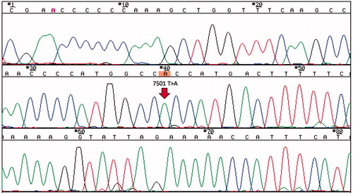
Figure 4. The B-mode echocardiogram. (A) The cardiac echogram on admission showed hypokinetic wall motion and anteroseptal hypokinesis. The ejection fraction determined by the modified Simpson’s method was 45.9%. (B) One year after the prescription of a β-blocker, the cardiac echogram showed normal wall motion of the anteroseptum. The ejection fraction determined by a modified Simpson’s method was 60.7%.
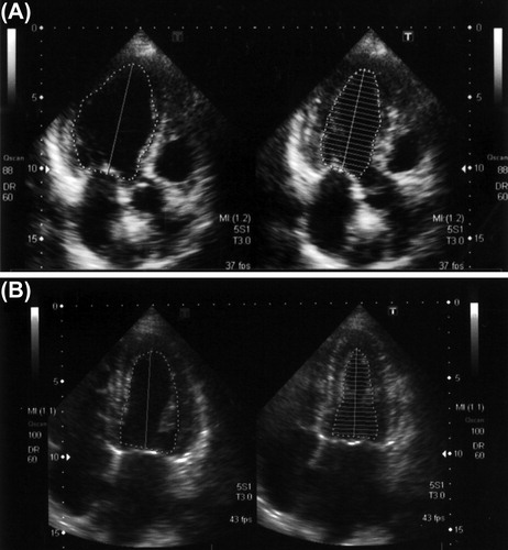
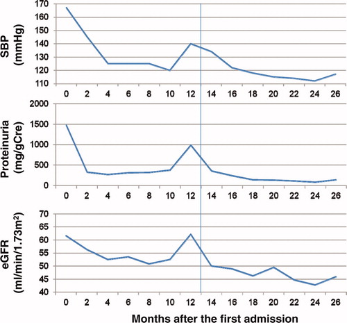
Figure 5. The changes of the systolic blood pressure (SBP), the degree of proteinuria and the estimated glomerular filtration rate (eGFR). The vertical line shows the time when the β-blocker was prescribed.