Figures & data
Table 1. Demographic date and clinical characteristics of patients.
Figure 1. Example of the glomerulitis and immunostaining for two markers (×400, DAB stain × 400): (A) glomerulitis with HE stain (arrow); (B) CD3-positive controls; (C) CD3-negative controls; (D) CD3-positive T lymphocytes in the glomerulus (arrow); (E) glomerulitis with PAS stain (arrow); (F) CD68-positive controls. (G) CD68-negative controls; (H) CD68-positive T lymphocytes in the glomerulus (arrow).
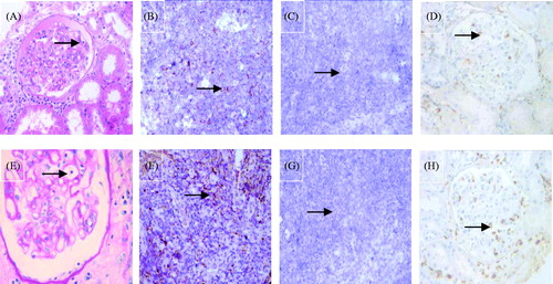
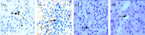
Figure 2. Example of the two PTCitis scores immunostaining for two markers (PAS stain × 1000, DAB stain × 1000). (A) Capillaritis with a PTCitis-score 1, i.e., max 3–4 luminal inflammatory cells in PTC (arrow). (B) Capillaritis with a PTCitis-score 2, i.e., max 5–10 luminal inflammatory cells in PTC (arrow). (C) CD3-positive T lymphocytes in the PTC (arrow). (D) CD68-positive macrophages in the PTC (arrow).
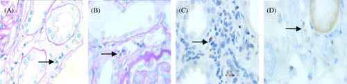
Figure 3. Immunostaining for C4d (DAB stain × 1000). (A) Negative C4d stain in PTC (arrow). (B) Minimal C4d stain/positive in PTC (arrow), i.e., C4d deposition in < 10% PTCs. (C) Focal C4d stain/positive in PTC (arrow), i.e., C4d deposition in 10–50% PTCs. (D) Diffuse C4d stain/positive in PTC (arrow), i.e., C4d deposition in > 50% PTCs.
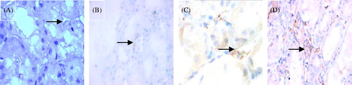
Table 2. Quantitative analysis of biopsy findings for C4d+ and C4d− groups.
Figure 4. Immunostaining for granzyme-B (DAB stain × 1000). (A) Granzyme B-positive controls (arrow). (B) Granzyme B-negative controls (arrow). (C) Granzyme B-positive T lymphocytes in the glomerulus (arrow). (D) Granzyme B-positive T lymphocytes in the PTC (arrow).
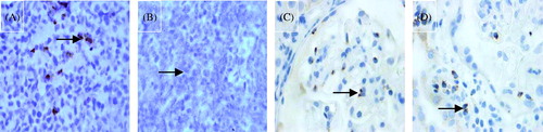
Figure 5. Immunostaining for Foxp3 (DAB stain × 1000). (A) Foxp3-positive controls. (B) Foxp3-negative controls (arrow). (C) Foxp3-negative T lymphocytes in the glomerulus (arrow). (D) Foxp3-negative T lymphocytes in the PTC (arrow).