Figures & data
Table 1. Coefficient variation of retrobulbar blood flow and RI of study population.
Table 2. Demographic properties.
Table 3. Biochemical assessment.
Table 4. Echocardiographic parameters.
Figure 1. The comparison of CIMT of patients with AA amyloidosis to healthy volunteers.
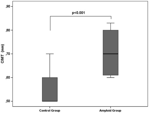
Figure 2. The correlation between Hs-CRP and CIMT of the study population.
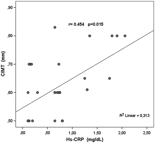
Figure 3. The comparison of OARI of patients with AA amyloidosis to healthy volunteers.
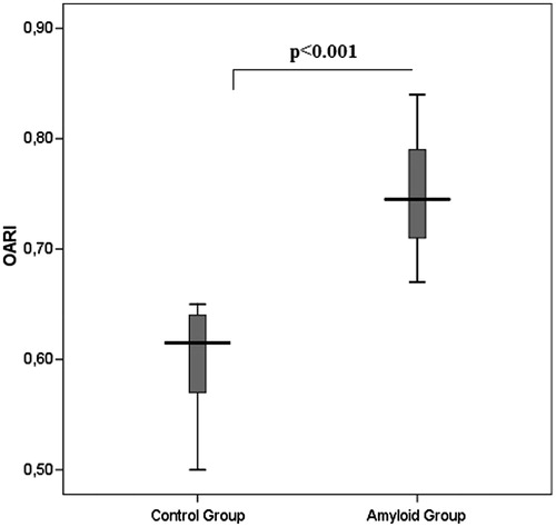
Table 5. Retrobulbar blood flow velocities.
Figure 4. The correlation between OARI and CIMT of the study population.
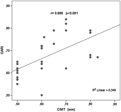
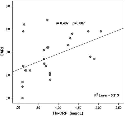
Figure 5. The correlation between Hs-CRP and OARI of the study population.