Figures & data
Table 1. Renal function parameters in the study groups (x ± s).
Figure 1. Effect of contrast media on apoptosis-related protein caspase-3 activation. Representative immunohistochemical findings (magnification 400×) of caspase-3 in normal rat kidney (A) and diabetic rat kidney (B) after normal saline (NS) or meglumine diatrizoate (DTZ) treatment. The meglumine diatrizoate induced increased caspase-3 expression in normal kidney, but the difference did not reach statistical significance (A,C). Especially in the diabetic kidney, the expression of caspase-3 was also significantly increased after intravenous injection of DTZ compared with normal saline (p < 0.05) (B,C). IOD = integrated optical density #p < 0.05 versus N group; *p < 0.05 versus NC group; §p < 0.05 versus D group.
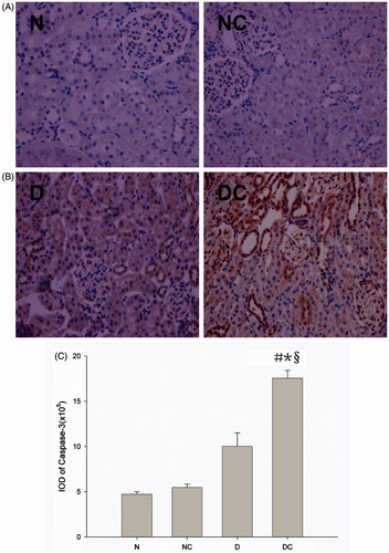
Figure 2. Effect of contrast media on Bcl-2 family proteins expression. Representative western blot findings of Bcl-2 (A) and Bax (B) in the kidney of each group. Diabetic rats had higher Bax and lower Bcl-2 expression than normal rats in the contrast media treated group (both p < 0.01) (C). The expression of Bcl-2 was also significantly decreased and the expression of Bax was significantly increased in the diabetic kidney after intravenous injection of meglumine diatrizoate compared with normal saline (both p < 0.05) (C). IOD = integrated optical density #p < 0.05 versus N group; *p < 0.05 versus NC group; §p < 0.05 versus D group.
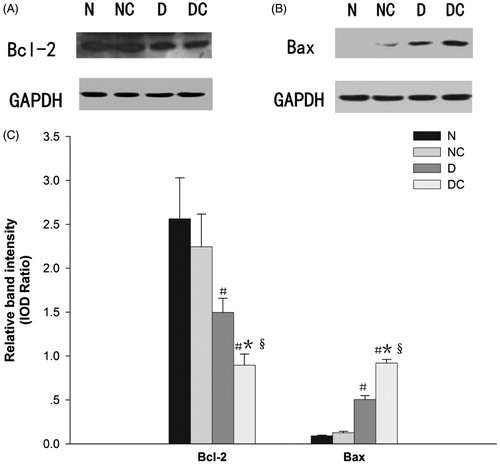
Figure 3. Effect of contrast media on phosphorylation of ERK1/2 and JNK. Representative western blot findings of p-ERK1/2 (A) and p-JNK (B) in the kidney of each group. The expression of p-JNK was significantly increased in the diabetic kidney after intravenous injection meglumine diatrizoate compared with normal saline (p < 0.05) (B,C), however, the expression of upstream signal molecule p-ERK1/2 was just the opposite (p < 0.05) (A,C). Moreover, the expression of upstream signal molecule p-JNK in diabetic kidney was significantly higher than that of normal rats in the contrast media-treated group (p < 0.05) (B,C). IOD = integrated optical density #p < 0.05 versus N group; *p < 0.05 versus NC group; §p < 0.05 versus D group.
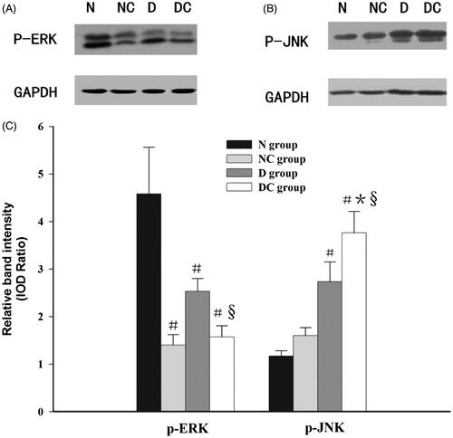
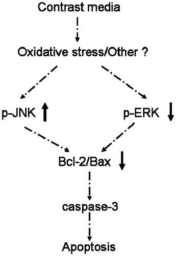
Figure 4. Pathophysiologic mechanisms for the apoptosis induced by ionic high-osmolar contrast media in diabetic rats.