Figures & data
Table 1. Oligonucleotide primer sets for real-time PCR.
Figure 1. IL-1α-induced fibroblast-like morphological changes in cultured NRK52E tubular epithelial cells. (a) Confluent cells cultured in medium alone for 4 days to maintain the cobblestone epithelial morphological state and growth pattern. (b) Cells co-cultured with IL-1α showed marked hypertrophy, became elongated, and developed an invasive growth pattern. (c) Cells grown in medium alone displayed a normal aggregated epithelial growth pattern with apical-basal polarity and numerous microvillus on the surface. (d) Cells co-cultured with IL-1α showed fibroblast-like morphological transition.
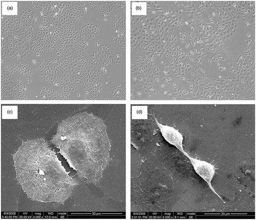
Figure 2. Real time PCR analysis of P311, α-SMA and TGF-β1 mRNA expression in all groups.
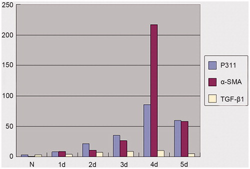
Table 2. Expression of P311, α-SMA, and TGF-β1 in all groups.
Figure 3. P311 protein and α-SMA protein in tubular epithelial-myofibroblast transition (TEMT). NRK52E cells were grown for four days on glass slides in the presence or absence of IL-1α or a neutralizing anti-P311 antibody. Cells expressing P311 protein or α-SMA protein were detected by immunohistochemistry. (a) Cells cultured in medium alone showed a normal epithelial morphology state. Only rare cells stained positive for P311. (b) Cells co-cultured with IL-1α caused marked TEMT. (c) The addition of neutralizing anti-P311 antibody produced much inhibition of P311 protein expression. (d) Cells cultured in medium alone showed a normal epithelial morphology and rare cells were stained positive for α-SMA. (e) Cells co-cultured with IL-1α caused marked TEMT. Strong α-SMA staining was demonstrated in many transformed cells. (f) The addition of neutralizing anti-P311 antibody obviously decreased the expression of α-SMA protein. This is one of three experiments that produced similar results.
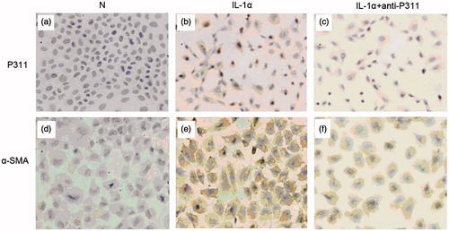
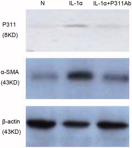
Figure 4. Western blot analyses of P311 and α-SMA protein in NRK52E cells. Cells were cultured in the presence of medium alone, IL-1α, or IL-1α plus a neutralizing anti-P311 antibody.