Figures & data
Table 1. Data of patients with complications after renal transplantation.
Figure 1. (a–c) An 18-year-old male with elevated serum creatinine level and uncontrolled hypertension 3 months post-operatively. (a) Angiography showed about 80% stenosis (arrowhead) at the anastomosis of transplant renal artery. (b) Balloon was dilated until no “waist-sign” (arrowhead) was left. (c) Subsequent angiography showed almost no residual stenosis (arrowhead).
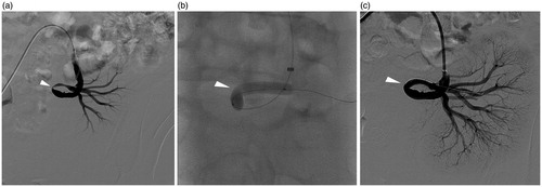
Figure 2. (a–d) A 33-year-old male with elevated serum creatinine level 6 days post-operatively. (a) Angiography showed about 80% stenosis (arrowhead) at the distal part of transplant renal artery near the renal hilum. (b) Balloon dilation was primary performed. Notice that the balloon diameter was selected in according with distal segmental artery (arrowhead). (c) Over 30% stenosis (arrowhead) was left after balloon dilation. (d) A balloon-expandable stent was deployed with almost no residual stenosis (arrowhead) left.
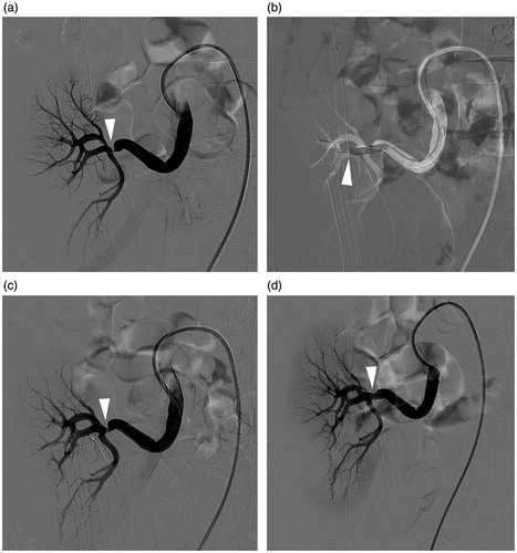
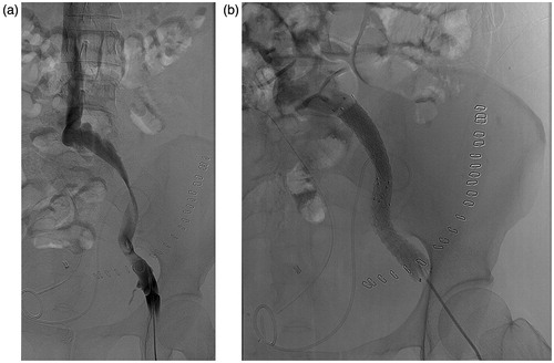
Figure 3. (a and b) A 44-year-old female with acute left leg swelling 14 days post-operatively. (a) Angiography showed extensive stenosis of the iliac vein caused by kinking. (b) Two bare stents in series were inserted and achieved perfect patency.