Figures & data
Figure 1. Light microscopy of renal cortical tissues in different groups for H&E. (a) In control, normal renal tissue morphology was seen. (b) After MTX, severe tubuler and glomeruler damage was noted. (c) QE treatment prevented tubuler and glomeruler damage compared with alone MTX group. (d) Renal injury degree was significantly decreased in the MTX + QE group when compared to MTX group. Asterisk: glomerular congestion and degeneration, Arrowhead: tubular cell swelling, Arrow: tubular dilatation. (H&E, scale bar, 50 μm). ap < 0.001 compared to control group, bp < 0.01 compared to MTX group.
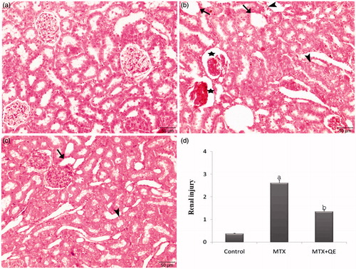
Figure 2. TUNEL staining of renal cortical tissues in different groups. (a) In control group, a few TUNEL positive cells were observed in the renal cortical tissues. (b) The TUNEL positive cells were significantly higher in the renal cortical tissues of the MTX treated group. (c) QE treatment markedly decreased the number of TUNEL positive cells. (d) The apoptotic index was significantly decreased in the MTX+QE group when compared to MTX group. (Arrow: TUNEL positive cells), (TUNEL, scale bar, 50 μm). ap < 0.001 compared to control group, bp < 0.01 compared to MTX group.
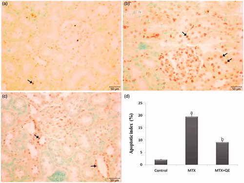
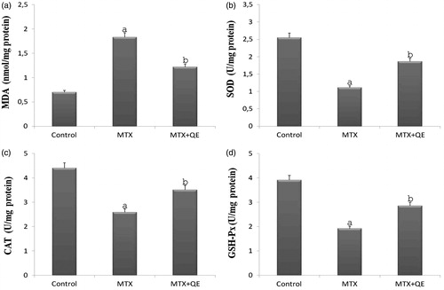
Figure 3. Renal tissue MDA, SOD, CAT, and GSH-Px levels in control, MTX, and MTX + QE groups. Tissue MDA levels were significantly increased, SOD, CAT, and GSH-Px levels were significantly decreased in MTX rats in comparison to control rats. Treatment of QE significantly decreased the elevated tissue MDA levels and increased of reduced SOD, CAT, and GSH-Px levels in the renal tissues. ap < 0.001 compared to control group, bp < 0.01 compared to MTX group.