Figures & data
Figure 1. Summary of the experimental design of experiments 1 and 2.
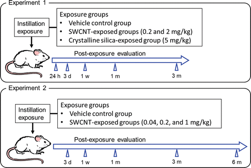
Table 1. Characterization of bulk SWCNTs and SWCNTs dispersed in the testing solution.
Figure 2. Morphology of the bulk SWCNTs and SWCNTs dispersed in the testing solution. Panels A–C show transmission electron microscopy (TEM) images of bulk SWCNTs at different magnifications. Panel d shows an atomic electron microscopy (AFM) image of SWCNTs dispersed in the testing solution. Panels e and f show TEM images of SWCNTs dispersed in the solution at different magnifications.
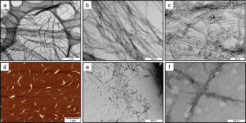
Figure 3. Distribution of SWCNT bundle diameter (left) and length (right) in 2 mg/mL SWCNT dispersion measured from digital images acquired by atomic force microscopy.
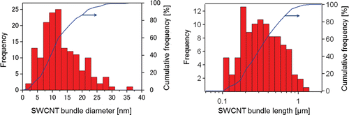
Figure 4. The resonance Raman scattering spectra of bulk SWCNT samples, and 0.2 and 2 mg/mL of SWCNT dispersions in the frequency regions of 100–3000 cm−1. The inset shows the spectra of the low frequency region. Abbreviations G and D denote G-band and D-band, respectively.
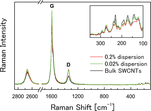
Figure 5. Absolute left lung weights of SWCNT-exposed rats and corresponding controls at each time point in experiment 1 (left) and 2 (right). Values are represented as the mean ± SD. *Significant increase from vehicle control (p < 0.05).
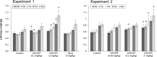
Figure 6. Number of neutrophils (a), macrophages (b), lymphocytes(c), and eosinophils (d) in BALF of SWCNT-exposed rats and corresponding controls at each time point in experiments 1 (left column) and 2 (right column). Values are represented as the mean ± SD. *Significant increase from vehicle control (p < 0.05).
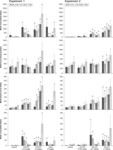
Figure 7. LDH value (a), protein content (b), and IL-1βactivity (c) in BALF of SWCNT-exposed rats and corresponding controls at each time point in experiments 1 (left column) and 2 (right column). Values are represented as the mean ± SD. *Significant increase from vehicle control (p < 0.05).
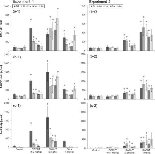
Table 2. Pulmonary histopathology severity scores* of rats in experiments 1 and 2.
Table 3. Summary of the results in experiments 1 and 2.
Figure 8. Light micrographs of lung tissue sections of rats at 1week after instillation in experiment 1 (H&E stain). No significant changes were observed in the vehicle control group (panel a). Minimal macrophage accumulation was observed in the alveoli of the 0.2 mg/kg SWCNT-exposed group (panel b). Moderate macrophage accumulation accompanied with foamy macrophages and mild inflammatory cell infiltration in the alveoli, and moderate macrophage infiltration and granuloma in the interstitium were observed in the 2.0 mg/kg SWCNT-exposed group (panel c). Minimal macrophage accumulation was observed in the alveoli of the crystalline silica-exposed group (panel d).
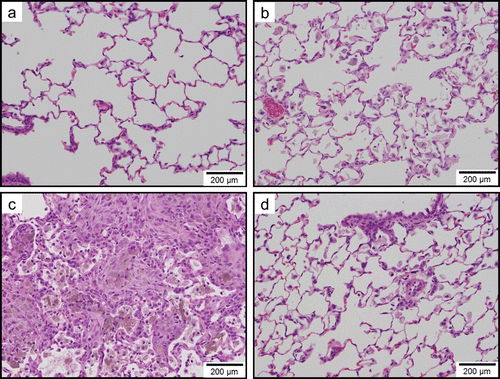
Figure 9. Light micrographs of lung tissue sections of rats at 3 months after instillation in experiment 1 (H&E stain). No significant changes were observed in the vehicle control group (panel a). Minimal macrophage accumulation was observed in the alveoli of the 0.2 mg/kg SWCNT-exposed group (panel b). Moderate macrophage accumulation accompanied with foamy macrophages, mild inflammatory cell infiltration in the alveoli, mild macrophage infiltration and granuloma in the interstitium, and mild hypertrophy of alveolar epithelium were observed in the 2.0 mg/kg SWCNT-exposed group (panel c). Mild macrophage accumulation accompanied with foamy macrophages, minimal inflammatory cell infiltration in the alveoli, and minimal hypertrophy of alveolar epithelium were observed in the crystalline silica-exposed group (panel d).
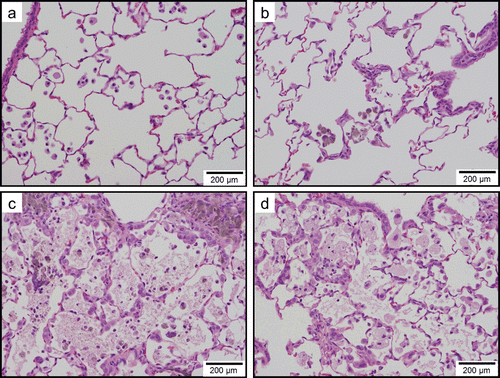
Figure 10. Light micrographs of lung tissue sections of rats at 6 months after instillation in experiment 2 (H&E stain). Minimal macrophage accumulation was observed in the alveoli of the vehicle control group. Minimal macrophage accumulation was observed in the alveoli of the 0.04 mg/kg SWCNT-exposed group (panel b). Minimal macrophage accumulation in the alveoli and interstitium were observed in the 0.2 mg/kg SWCNT-exposed group (panel c). Mild macrophage accumulation accompanied with foamy macrophages and minimal inflammatory cell infiltration, minimal macrophage infiltration in the interstitium, and minimal hypertrophy of alveolar epithelium were observed in the 1 mg/kg SWCNT-exposed group (panel d).
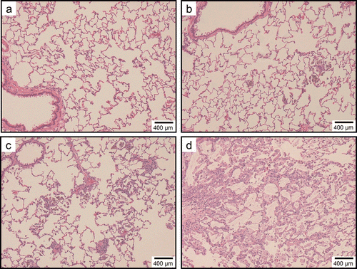
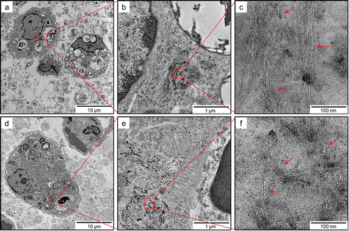
Figure 11. TEM images of SWCNTs deposited in lungs of rats exposed to 2.0 mg/kg SWCNTs at 1 month (panels a–c) and 3 months (panels d–f) after instillation at different magnifications. At any of the time points, SWCNTs deposited in the lungs were typically phagocytosed by alveolar macrophages or macrophages in the interstitial tissues.