Figures & data
Figure 1. Fundus photographs showing well-demarcated area of retinal whitening in the posterior pole with multiple cotton wool spots in both eyes and retinal hemorrhage in the left eye.
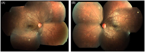
Figure 2. Fluorescein angiography pictures of right eye showing increased foveal avascular zone and multiple areas of patchy capillary dropouts in the periphery and posterior pole.
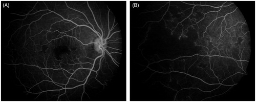
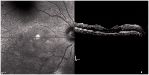
Figure 3. SD-OCT scan through fovea showing thickening of inner retinal layers and shadowing of outer retinal layers with a clear demarcation at the margin of widened foveal avascular zone (arrows). The ELM, IS-OS junction, and COST line were disrupted at the fovea.
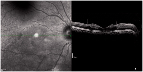
Figure 4. SD-OCT after 6 weeks. The ELM, IS-OS junction, and COST line started regaining their normal architecture but the inner retinal layers looked the same.