Figures & data
TABLE 1. Inclusion and exclusion criteria for participation in study.
TABLE 2. Demographics of enrolled patients.
TABLE 3. Parameters of enrolled patients at baseline compared to 26 weeks.
TABLE 4. The progression of parameters over 26 weeks.
FIGURE 1. Pattern 1 shows a progressive resolution of CME and significant improvement at 2 weeks with complete resolution after the second injection that persisted throughout the study. Pattern 2 shows a two-phase improvement: very fast initial improvement of CME noted at 2 days then reemergence of the fluid at 2 weeks with complete resolution after the second injection that persisted throughout the study.
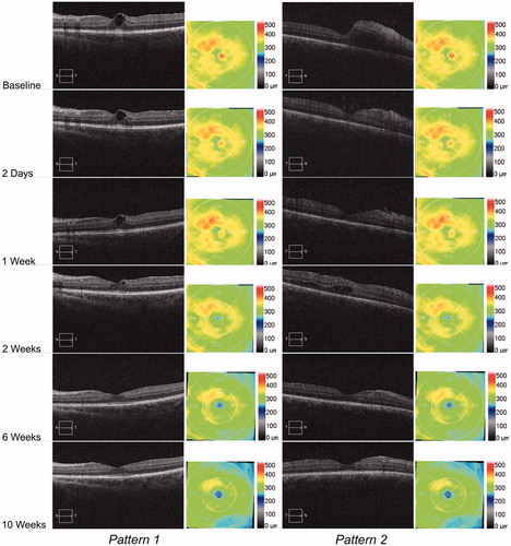
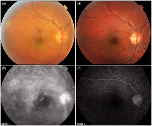
FIGURE 2. (A) Fundus photo of eye number 5 showing significant vitreous haze and inferior vitreous hemorrhage secondary to inferior neo-vessel. (B) Complete resolution of vitreous haze and inferior neo-vessel on last follow-up. (C) Late frame of fluorescein angiography of eye 5 showing diffuse optic disc hyperfluorescence, macular edema with pooling of dye in cystic spaces, diffuse retinal vascular staining, and diffuse capillary leakage in the posterior pole. (D) Complete resolution of leakage on last follow-up.