
Figures & data
FIGURE 1. Scleromalacia located in the upper area of the temporal bulbar quadrant. (a). Note the loss of scleral tissue centrally and a change of transparency in the adjacent tissue, visible as a bluish discoloration. (b). The same eye is shown 8 years later with marked progression of the scleromalacia and a bulging staphyloma.
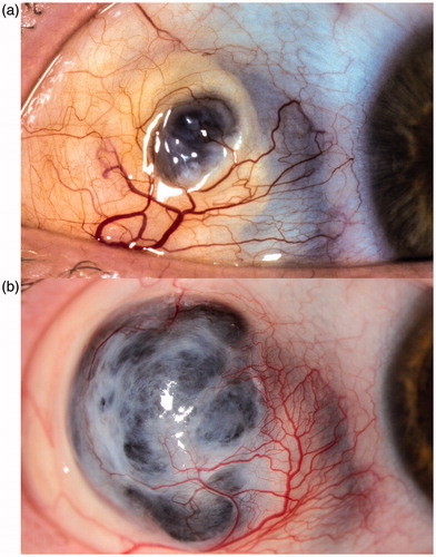
FIGURE 2. Fundoscopic photograph of the same eye with hemorrhagic occlusive vasculitis and confluent whitish areas of retinal necrosis in the peripheral ocular fundus. Note the central involvement with macular hemorrhage.
