Figures & data
Figure 1. Light microscopy of a healthy individual’s whole blood smear (scale = 10) (A); SEM micrograph of a typical discoid RBC (scale = 1 μm) (B); Typical bulbous platelet with some pseudopodia (scale = 1 μm) (C); Fibrin network (scale = 1 μm) (D).
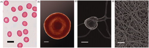
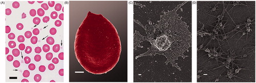
Figure 2. Light microscopy of a whole blood smear with arrows showing platelets between erythrocytes (scale = 10 μm) (A); Erythrocyte with changed ultrastructure (scale = 1 μm) (B); SEM micrograph of activated platelet with spreading and membrane changes (scale = 2 μm) (C); Numerous platelets within dense fibrin networks (scale = 2 μm) (D).