Figures & data
Table I. Irradiation conditions for each intercomparison.
Table II. Summary of exercise participation.
Figure 1. Intercomparisons across laboratories of estimated doses obtained using (A) conventional dicentric chromosome assay (CDCA) analysis of 50 metaphase spreads, (B) QuickScan DCA of 50 metaphase spreads, and (C) cytokinesis block micronucleus (CBMN) analysis of 200 binucleated cells. Each data point is a dose estimate from one individual, with scorers from each laboratory shown using the same symbol. The solid lines represent ± 0.5 Gy intervals.
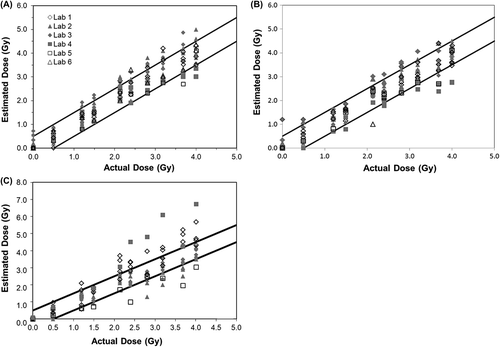
Figure 2. Illustration of the percentage of samples with dose estimates in agreement between laboratories for each year for each endpoint. A tally was prepared of the samples in which all laboratories were in agreement of the dose estimate (p > 0.05). The average dose estimate from each laboratory and method was calculated and compared using ANOVA.
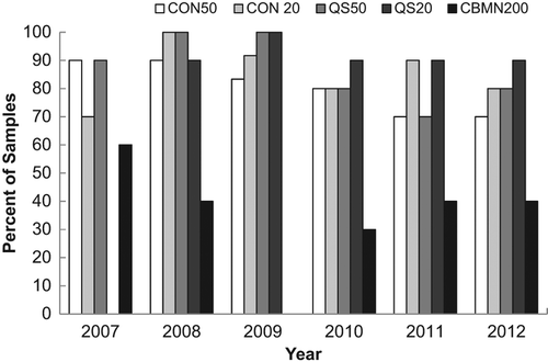
Table III. Sample ANOVA analysis of data from year 2008; dose delivered = 1.8 Gy.
Figure 3. Illustration of the percentage of samples with dose estimates (A) within 0.5 Gy of the given dose, (B) more than 0.5 Gy over the given dose, and (C) more than 0.5 Gy under the given dose.
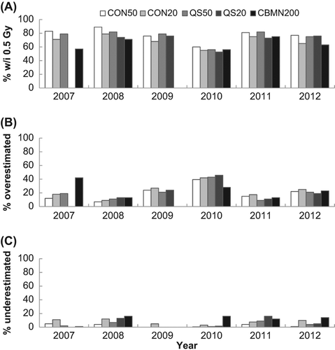
Figure 4. Illustration of the average time required to score one sample for each method.
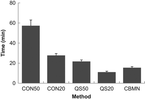
Table IV. Performance statistics z sample analysis of data from year 2008; dose delivered = 1.8 Gy.
Figure 5. A comparison of the percentage of correctly evaluated samples based on a |z| ≤ 2 for each method across all years of the intercomparisons.
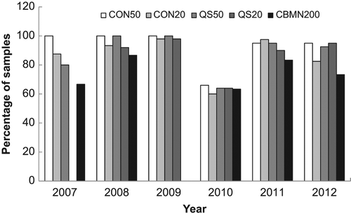
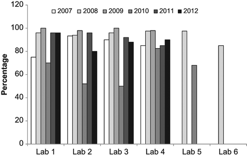
Figure 6. A comparison of the percentage of correctly evaluated samples based on a |z| ≤ 2 for each method in each laboratory.
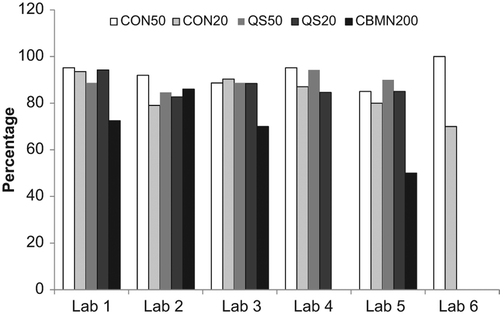
Figure 7. A comparison of the percentage of correctly evaluated samples based on a |z| ≤ 2 for each year in each laboratory.