1,803
Views
32
CrossRef citations to date
0
Altmetric
Research Article

An emerging consensus on aquaporin translocation as a regulatory mechanism

, &
Pages 101-112 | Received 21 Aug 2012, Accepted 09 Oct 2012, Published online: 21 Nov 2012

Figures & data

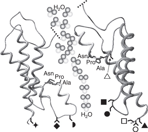
Table I. Tissue distribution and functional roles of the 13 human AQPs. The Table summarizes the distribution and function of AQPs in specific cells and tissues of the human body. Immunohistochemistry has revealed the diversity of AQP distribution within cells and tissues. For example, AQPs 1, 2, 3, 4, 6, 7, 8, 9, 10 and 11 have all been shown to be expressed in tissues of the kidney whereas AQP0 is almost exclusively expressed in lens epithelial cells. AQP11 expression in leucocytes may explain the immunohistochemical identification of AQP11 in other tissues.

Figure 1. Schematic diagram of the shared structural architecture of aquaporins. Six transmembrane α-helices are connected by alternating extracellular and intracellular loops with the carboxyl and amino termini orientated towards the cytoplasm. Two of these connecting loops (B and E) fold into the transmembrane pore so that the protein resembles an hour-glass. Loops B and E each form a helical region containing highly conserved Asn-Pro-Ala (NPA) motifs that facilitate selectivity of the pore. The approximate positions of serine or threonine protein kinase phosphorylation sites involved in AQP translocation are indicated as follows; ▴ = Ser235 in AQP0; ⧫ = Thr157 and ○ = Thr239 in AQP1; □ = Ser256 in AQP2; = Ser111 and = Ser180 in AQP4; = Ser152 in AQP5; = Ser11 and ▵ = Ser222 in AQP9.

Figure 1. Schematic diagram of the shared structural architecture of aquaporins. Six transmembrane α-helices are connected by alternating extracellular and intracellular loops with the carboxyl and amino termini orientated towards the cytoplasm. Two of these connecting loops (B and E) fold into the transmembrane pore so that the protein resembles an hour-glass. Loops B and E each form a helical region containing highly conserved Asn-Pro-Ala (NPA) motifs that facilitate selectivity of the pore. The approximate positions of serine or threonine protein kinase phosphorylation sites involved in AQP translocation are indicated as follows; ▴ = Ser235 in AQP0; ⧫ = Thr157 and ○ = Thr239 in AQP1; □ = Ser256 in AQP2; = Ser111 and = Ser180 in AQP4; = Ser152 in AQP5; = Ser11 and ▵ = Ser222 in AQP9.

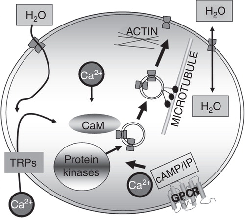
Table II. Triggers of aquaporin translocation to the plasma membrane (PM). The Table summarizes known stimuli of the dynamic translocation of AQPs, where possible avoiding observations solely linked to water permeability (WP) and/or transcription/translation. The experimental system used to determine the AQP translocation is included for each trigger.

Table III. Intracellular components involved in the translocation of specific aquaporins to the plasma membrane (PM). The Table summarizes the components of dynamic translocation of AQPs avoiding, where possible, observations solely linked to water permeability (WP) and/or transcription/translation. Arrows indicate the direction of the effect. The experimental system used to determine the AQP translocation is included for each trigger and the residue that the component acts upon is also included where known. †Residues in PKC. *AQP2 is not added in more detail than PKA-linked as this has been very well reviewed elsewhere (Valenti et al. Citation2005).

Figure 2. Trigger-induced aquaporin translocation: a regulatory mechanism for cellular water flow. Some or all of the components shown may be involved in the translocation of different AQPs. For example, AQP1 translocation is known to be triggered by hypotonicity, which causes calcium influx through transient receptor potential (TRP) channels and subsequent calmodulin-mediated PKC phosphorylation of specific AQP1 threonine residues, resulting in microtubule-dependent AQP1 translocation. Other protein kinases are thought to be involved in AQP translocation such as PKA-mediated translocation of AQP2 and AQP5 following activation of vasopressin V2 and M3 muscarinic GPCRs, respectively.

Figure 2. Trigger-induced aquaporin translocation: a regulatory mechanism for cellular water flow. Some or all of the components shown may be involved in the translocation of different AQPs. For example, AQP1 translocation is known to be triggered by hypotonicity, which causes calcium influx through transient receptor potential (TRP) channels and subsequent calmodulin-mediated PKC phosphorylation of specific AQP1 threonine residues, resulting in microtubule-dependent AQP1 translocation. Other protein kinases are thought to be involved in AQP translocation such as PKA-mediated translocation of AQP2 and AQP5 following activation of vasopressin V2 and M3 muscarinic GPCRs, respectively.

Reprints and Corporate Permissions

Please note: Selecting permissions does not provide access to the full text of the article, please see our help page How do I view content?

To request a reprint or corporate permissions for this article, please click on the relevant link below:

Academic Permissions

Please note: Selecting permissions does not provide access to the full text of the article, please see our help page How do I view content?

Obtain permissions instantly via Rightslink by clicking on the button below:

If you are unable to obtain permissions via Rightslink, please complete and submit this Permissions form. For more information, please visit our Permissions help page.