Figures & data
Figure 1. Example trace showing the change in fluorescence upon mixing (arrow) of 100 µg/ml sodium metasilicate and FPE labelled HOBs with the single exponential fit. This Figure is reproduced in colour in the online version of Molecular Membrane Biology.
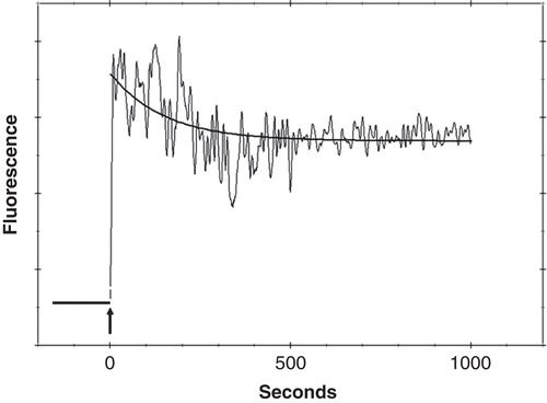
Figure 2. FPE labelled osteoblast imaged using a Leica SP2 Confocal Laser Scanning Microscope. (A) shows a FPE labelled osteoblast – fluorescence can be clearly identified at the cell membrane. (B) shows the same cell, after the addition of 100 µg/ml sodium metasilicate. A decrease in fluorescence is observed. (C) shows the same cell, after the addition of Ca2+ ions (5 mM CaCl2). An increase in fluorescence is clearly visible. This Figure is reproduced in colour in the online version of Molecular Membrane Biology.
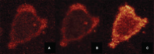
Figure 3. Binding profile based on a complete set of sodium metasilicate titrations with FPE labelled human osteoblasts. The black line shows the fit for a hyperbolic function (Equation 2). The dashed line shows the fit for a sigmoidal function (Equation 3). This Figure is reproduced in colour in the online version of Molecular Membrane Biology.
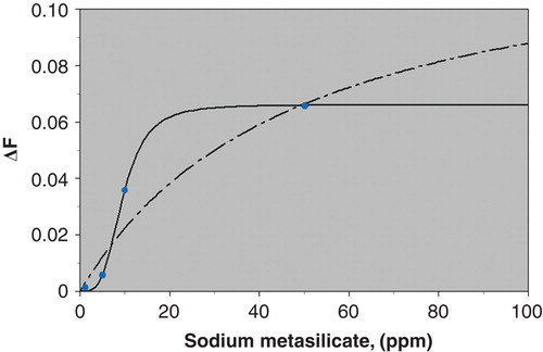
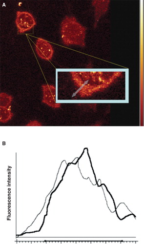
Figure 4. Human osteoblasts labelled with FPE and imaged using a Leica SP2 Confocal Laser Scanning Microscope. Fluorescence can be clearly identified at the cell membrane. The region of interest for analysis is identified by the arrow across the cell membrane (A). Intensity profiles were generated by measuring the fluorescence intensity across the ROI before (black) and after (grey) the addition of sodium metasilicate (B). This Figure is reproduced in colour in the online version of Molecular Membrane Biology.