
Figures & data
Figure 1. The BBB and the neurovascular unit. The blood brain barrier consist of a modified endothelium, which overexpresses tight junctions and Adherens junctions, surrounded by pericytes, astrocytical processes and neurons.
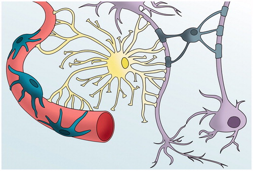
Figure 2. The tight junctions. Structure of the main proteins that form the tight junctions. They are proteins with extracellular domains that mediate physical interactions and intracellular domains that anchor to the cytoskeleton.
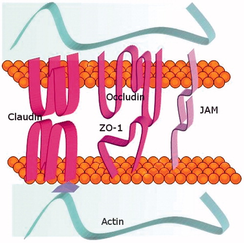
Table 1. Tight junction proteins.
Figure 3. Physiological crossing of the BBB. Representation of the paracellular route and transcellular routes such as carrier-mediated endocytosis, efflux pumps, receptor-mediated endocytosis and adsorptive-mediated transcytosis.
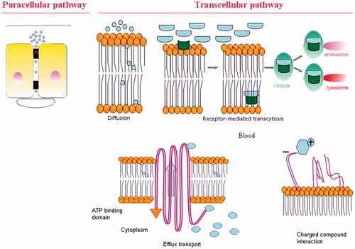
Figure 4. A schematic representation of current strategies to deliver drugs to the brain by invasive techniques. It encloses surgery-needed approaches and BBB disruption whilst non-invasive techniques include drug modification by medicinal chemistry approaches and drug encapsulation through nanotechnological carriers.
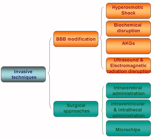
Figure 5. A schematic representation of current strategies to deliver drugs to the brain by non-invasive techniques. Non-invasive techniques include drug modification by medicinal chemistry approaches and drug encapsulation through nanotechnological carriers.
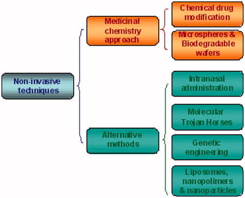
Figure 6. Schematic representation of the three different types of liposomes. Small Unilamellar Vesicles (SUV), Large Unilamellar Vesicles (LUV) and Multilamellar (MLV).
