Figures & data
Figure 1. Impairment of corticosterone response to restraint stress and open-field exposure in PACAP− / − mice. After restraint (A), open-field exposure (B), cold exposure (C), or ether-inhalation (D) stress, plasma corticosterone levels were determined in PACAP− / − (closed symbols) and wild-type (open symbols) mice (n = 3–8 per group). **P < 0.01 compared with time 0, except for (C), 22°C, in the same genotype; ##P < 0.01 compared with wild-type mice.
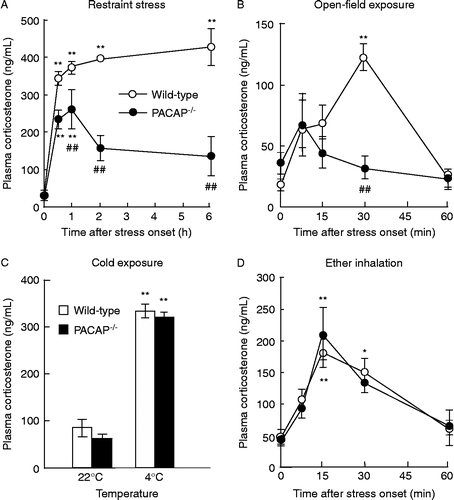
Figure 2. CRF- and ACTH-induced increase in plasma corticosterone levels in PACAP− / − mice. Thirty minutes after injection of CRF (A) or ACTH (B), the plasma corticosterone levels were determined in PACAP− / − (closed circles) and wild-type (open circles) mice (n = 3–6 per group). **P < 0.01 compared with 0 μg/kg.
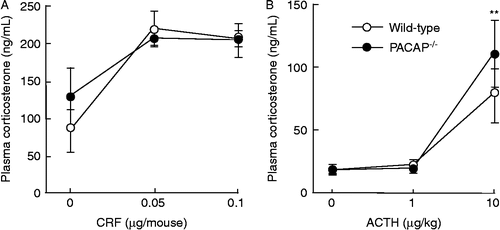
Figure 3. Marked attenuation of restraint stress-induced c-Fos expression in CRF-positive PVN neurons in PACAP− / − mice. PACAP− / − and wild-type mice with and without restraint stress were subjected to double immunofluorescence staining for c-Fos plus CRF (A, C, and D) or AVP (B, C, and E). (A and B) Representative microscope images are shown. Insets, higher magnification images of the regions boxed. Arrowheads indicate double-positive cells. Scale bars, 50 μm. (C–E) The total number of c-Fos-positive cells (C), the fraction of CRH neurons that was c-Fos positive (D), and the fraction of AVP neurons that was c-Fos positive (E) were analyzed in PACAP− / − (closed bars) and wild-type (open bars) mice (n = 6–7 per group). **P < 0.01.
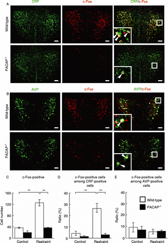
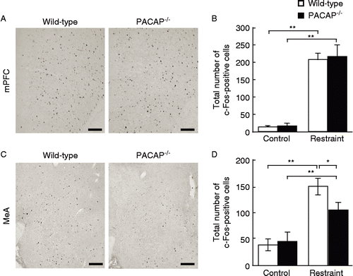
Figure 4. Restraint stress-induced c-Fos expression in the mPFC and MeA in PACAP− / − mice. PACAP− / − and wild-type mice with and without restraint stress were subjected to immunostaining to detect c-Fos expression in the mPFC (A and B) and MeA (C and D). (A and C) Representative microscope images are shown. Scale bars, 100 μm. (B and D) The quantitative results in PACAP− / − (closed bars) and wild-type (open bars) mice are shown (n = 6–7 per group). *P < 0.05; **P < 0.01.