Figures & data
Table I. UCMS protocol.
Figure 1. Effects of chronic psychological stress on (A) frequency of grooming, (B) food intake, and (C) body weight gain in rabbits. Grooming frequency was measured by counting the number of attempts made to clean the fur by licking or scratching for 5 min after spraying a 10% sucrose solution on the dorsal coat. Data are mean ± SEM. *P < 0.01 vs. control group, two-way ANOVA (n = 9–10 per group/time point).
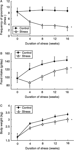
Figure 2. Effects of chronic psychological stress on serum corticosterone concentrations. Data are mean ± SEM. *P < 0.01 versus control group, two-way ANOVA (n = 9–10 per group/time point).
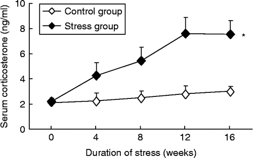
Table II. Effects of psychological stress on systemic hemodynamic parameters.
Figure 3. Expression levels of mRNAs for TNF-α, CRP, MCP-1, and macrophage MIF in the abdominal aorta in control and chronic variable stress groups measured by real-time PCR. Data are mean ± SEM. *P < 0.01 vs. control group, two-way ANOVA (n = 4–5 per group/time point).
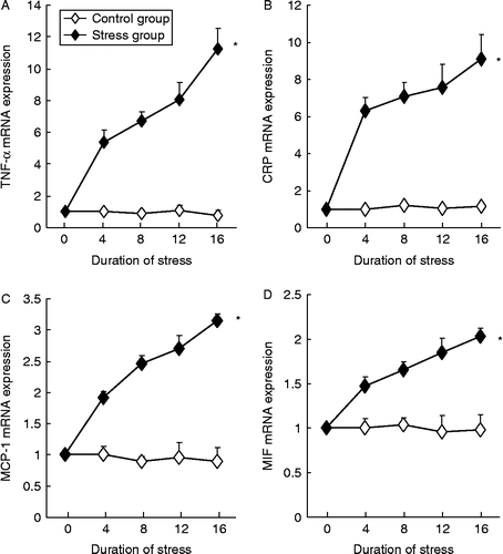
Figure 4. Chronic psychological stress increased the protein expression levels of TNF-α, CRP, ICAM-1, MCP-1, and macrophage MIF in the abdominal aorta. Antigens were detected with immunohistochemistry and visualized with DAB chromogen (brown color, arrows). Samples from the 4- and 12-week group showed a similar trend of upregulation of these inflammatory molecules (images not shown). Scale bar = 50 μm. The quantitative data for all treatment groups are shown as bar graphs on the right. Digital images were analyzed with Image-Pro Plus 6.0 software and results expressed as percentage of positively stained area. Data are mean ± SEM. *P < 0.05 vs. control (C, 16-week untreated animals), one-way ANOVA, n = 5 per group/time point.
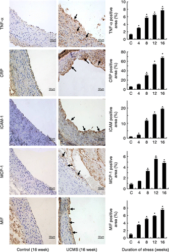
Figure 5. Macrophage infiltration and lipid accumulation in the subendothelial space at 16-week poststress exposure. Macrophages were identified by the anti-rabbit macrophage (RAM1) antibody (arrow); lipids were detected with Oil Red O staining (arrow). The sections were counterstained with hematoxylin. Scale bar = 50 μm.
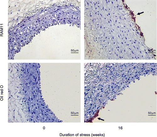
Figure 6. Effects of chronic psychological stress on serum TNF-α (A) and CRP (B) in control and stressed animals. Data are mean ± SEM. *P < 0.01 vs. control group, two-way ANOVA (n = 9–10 per group/time point).
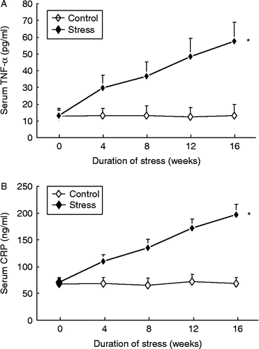
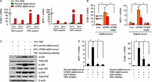
Figure 7. Serum from rabbits exposed to UCMS-treated rabbits increased MCP-1 and ICAM-1 expression in cultured murine vascular smooth muscle cells via TNF-α and p38/JNK pathways. (A) Real-time PCR results showing that MCP-1 and ICAM-1 mRNAs were upregulated by incubation with serum from stressed rabbits in a dose-dependent manner, as compared with serum from untreated rabbits (averaged data from two experiments). FBS, fetal bovine serum. (B) Serum-induced upregulation of ICAM-1 and MCP-1 mRNAs was blunted by pretreatment with neutralizing antibody against TNF-α (NAb, 2 μg/ml; data expressed as fold of those from cells maintained in 10% FBS), n = 3. (C) Western blots showing that stimulation with 20% serum from stressed rabbits increased phosphorylation of p38 and JNK, but not ERK, as compared with cells treated with serum from control rabbits. Phosphorylation of p38 and JNK was suppressed by TNF-α neutralizing antibody (example from three independent experiments). (D) Treatment with specific p38 and JNK inhibitors SB202190 (1 μM) and SP600125 (1 μM) significantly inhibited MCP-1 and ICAM-1 mRNA expression stimulated by serum from stressed rabbits. Data are mean (A) or mean ± SEM (B, D). *P < 0.05, one-way ANOVA (n = 3).