Figures & data
Table I. Thermal responses of TOL, MT, and INT mice in two heat tests.
Figure 1. Representative tracings of SBP and DBP (top), HR (middle), and Tc (bottom) in a conscious INT mouse before, during, and after heat exposure. The signals were recorded simultaneously in real time from a single experiment. The ambient temperature (Ta) was obtained inside a mouse cage.

Figure 2. Comparison of MAP and HR at baseline, during heat exposure, and recovery in TOL, MT, and INT mice. Peak and recovery values were obtained when Tc reached the highest and lowest levels during and after heat exposure, respectively. *P < 0.05, compared to TOL.
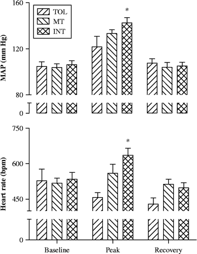
Table II. Plasma cytokines, 8-isoprostane, corticosterone, and SOD in control, TOL, MT, and INT mice.
Figure 3. Tissue HSP72 levels in control mice and in TOL, MT, and INT mice following heat stress. One-way ANOVA revealed significant (F (3, 37) = 4.9, 16.3, 8.7, 11.6, and 27; P = 0.006, 0.0001, 0.0002, 0.0001, and 0.0001 for liver, kidney, heart, gastrocnemius muscle, and soleus muscle, respectively). P < 0.05: *compared to control; †compared to TOL; #compared to MT.
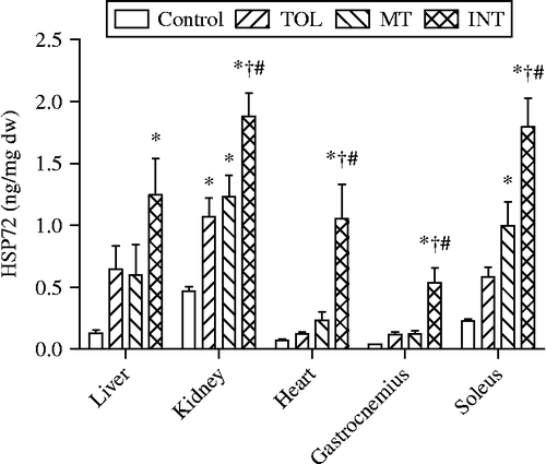
Figure 4. HSP90, GR, and HSF1 protein levels in liver, heart, and gastrocnemius muscle tissues of control mice and TOL and INT mice following heat stress. *P < 0.05, compared to control and TOL.
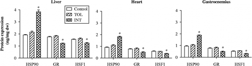
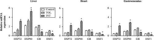
Figure 5. HSP72, HSP90, GR, and HSF1 mRNA levels in liver, heart, and gastrocnemius muscle tissues of control mice and TOL and INT mice following heat stress. *P < 0.05, compared to control and TOL.