Figures & data
Figure 1. Experimental groups showing period of stress exposure or recovery. Blood and tissue samples were collected at autopsy.
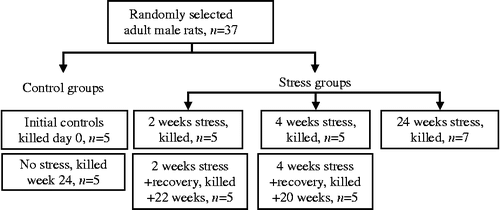
Figure 2. Vertical bars are mean ± SEM. Percentage change in body weight compared to initial body weight (A) and relative adrenal weight (B) in control, stressed, and recovery groups, n = 5 rats per group. *p < 0.05 versus controls, ANOVA, and Duncan's test.
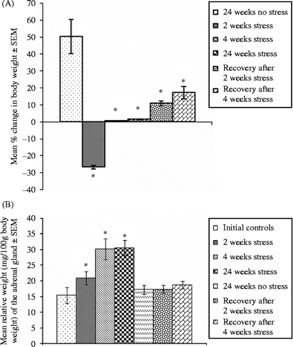
Table I. Stress and adrenal 3β-HSDH activity and plasma lipid profile.
Table II. Stress and activities of hepatic antioxidant enzymes and GGT.
Table III. Stress and aminotransferases and LDH activities.
Table IV. Stress and lipid peroxidation.
Figure 3. Transverse sections of similar areas of thoracic aorta of a control rat (24 weeks no stress; A and B), and a rat stressed for 24 weeks (C and D) and a part of the aorta of a stressed rat under high magnification to show a portion of an atherosclerotic plaque (E) and foam cells (arrow). Note the thickening of wall of the aorta and a fibrous layer in the vessel of the stressed rat. B and D are higher magnification images of A and C, respectively. Hematoxylin–eosin stain; AD, adventitia; EN, endothelium; FB, fibrous layer; FC, foam cells; ME, media.
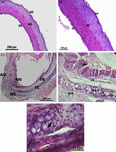
Figure 4. Transverse sections of the thoracic aorta of a control rat (24 weeks no stress; A), and a rat stressed for 24 weeks (B) stained with Oil Red O for localization of lipids. Note the Oil-Red-O-positive lipid deposition (arrow heads) in the section from a stressed rat (B). Transverse sections of the thoracic aorta of a control rat (24 weeks no stress; C), and a rat stressed for 24 weeks (D) stained with van Gieson technique to demonstrate elastic fibers (arrow head) and collagen (solid arrow). Note the presence of abundant elastic fibers in the control rat aorta (C) and depletion of elastic fibers and abundant collagen deposition in the stressed rat aorta (D).
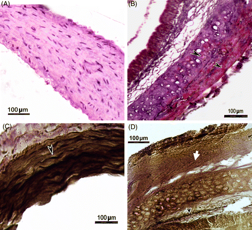
Table V. Stress and thickness of layers of thoracic aorta wall.