
Figures & data
Figure 1. The DxH recombinant inbred mouse strains both have a DBA/2J background, but the high-fear sensitized startle (H-FSS) strain has an insertion of four chromosomal C3H/2JHd segments into the DBA/2JHd background compared to the low (L)-FSS strain. (A) Increase in freezing duration following acclimation (0–2 min), and extinction learning during the first versus last 10 min of presentation of conditioned acoustic stimuli (CS). *p < 0.05 for fear recall (acclimation versus 3–12 min) and extinction learning (3–12 min versus 13–22 min), ^p < 0.05 for strain differences, &p < 0.05 strain x fear recall interaction. (B) H-FSS mice also show a severe extinction deficit in their freezing response. #p < 0.05 for strain differences. (C) Brief freezing in individual L-FSS mice upon CS presentation that declines rapidly, whereas H-FSS mice show generalized fear responses during acclimation and a deficit in extinction learning.
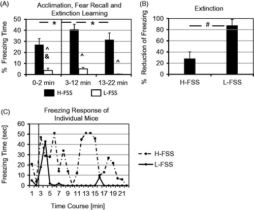
Figure 2. Immobility in the acute-15 min variant of the forced swim test (FSTv) and endocrine changes 30 min after exposure to acute swim stress (15 min duration). (A) The more fearful H-FSS mice spent a longer time immobile (i.e. floating) than the less fearful L-FSS mice in the acute FSTv. (B) There were no significant differences between basal plasma corticosterone concentrations of H-FSS and L-FSS mice, but acute swim stress induced by exposure to the FSTv resulted in a significant increase in corticosterone levels in both strains. L-FSS mice showed a higher stress-induced increase in plasma corticosterone than H-FSS mice (% of control), but this difference was not significant at the time point investigated after exposure to acute swim stress. *p < 0.05.
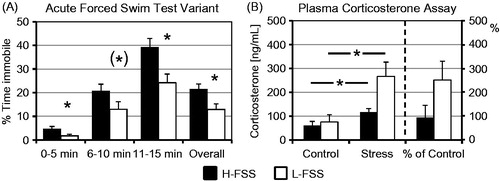
Figure 3. Basal noradrenaline levels and changes in noradrenaline 30 min after exposure to acute swim stress in the FSTv. The H-FSS mice display higher noradrenaline concentrations in the amygdala (A), medial prefrontal cortex (C), and hypothalamus (E), and lower noradrenaline concentrations in the striatum (D) than L-FSS mice, but the two mouse strains do not differ in hippocampal noradrenaline levels (B). A stress x strain interaction is seen in the hypothalamus (E), and a stress effect in the brainstem (F).∼p < 0.05 for stress effect, *p < 0.05 for strain effect and ^p < 0.05 for stress x strain interaction in the two-way ANOVA.
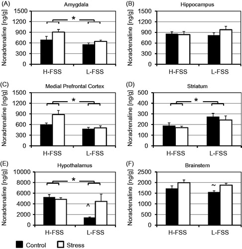
Figure 4. Basal dopamine levels and changes in dopamine 30 min after exposure to acute swim stress in the FSTv. Dopamine levels are higher in the amygdala (A), hypothalamus (E), and brainstem (F) and lower in the striatum (D) of H-FSS than L-FSS mice. No differences are observed in the hippocampus (B). The stress-induced increase in dopamine levels is significant in the striatum (D) and brainstem (F), and an additional stress x strain interaction is seen in the medial prefrontal cortex (C) and striatum (D). ∼p < 0.05 for stress effect, *p < 0.05 for strain effect, and ^p < 0.05 for stress x strain interaction in the two-way ANOVA.
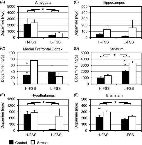
Figure 5. Basal serotonin levels and changes in serotonin 30 min after exposure to acute swim stress in the FSTv. Serotonin levels are higher in the amygdala (A) and hypothalamus (F) and lower in the striatum (D) of H-FSS than L-FSS mice. As with dopamine, no differences are observed in the hippocampus (B) and medial prefrontal cortex (C). The stress-induced increase in serotonin levels is significant in the amygdala (A) and brainstem (F), and a stress x strain interaction is seen in the hypothalamus (E). ∼p < 0.05 for stress effect, *p < 0.05 for strain effect, and ^p < 0.05 for stress x strain interaction in the two-way ANOVA.
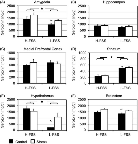
Table 1. Measurements of the dopamine metabolites 3,4-dihydroxyphenylacetic acid (DOPAC) and homovanillic acid (HVA) as well as the serotonin metabolite 5-hydroxyindoleacetic acid (5-HIAA) show significant differences in several brain regions in the two-way ANOVA.
Table 2. Dopamine turnover calculated as the ratio of the metabolites DOPAC and HVA to dopamine (DA) and serotonin turnover as the ratio of 5-HIAA to serotonin (5-HT). Strain differences and stress significantly influence dopamine and serotonin turnover in several brain regions as indicated by the two-way ANOVA.