Figures & data
Figure 1. Chemical structures of folpet and captan and their reaction products following interaction with thiols.
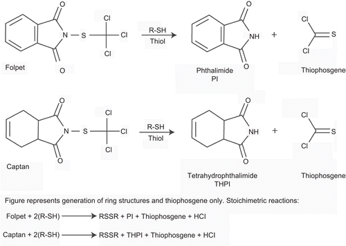
Figure 2. Reactions of folpet and thiophosgene with cysteine, a representative thiol, producing the urinary metabolite TTCA.
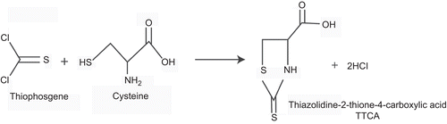
Table 1. Tumor incidences of selective tissues in a study in CD-1 mice administered folpet in the diet (CitationWong, 1985).
Table 2. Tumor incidences of selective tissues in a study in B6C3F1 mice administered folpet in the diet (CitationRubin and Nyska, 1985).
Figure 3. Opened gastric and duodenal lumen of high-dose group mouse. A large mass (squamous cell carcinoma) growing in the nonglandular portion of the stomach is indicated by a large asterisk. The duodenal lumen is obstructed by a large nodule (small asterisk). (Reproduced by permission from A. Nyska et al., Induction of gastrointestinal tumors in mice fed the fungicide folpet: possible mechanisms. 1990. Jpn J Cancer Res 81:545–549.)
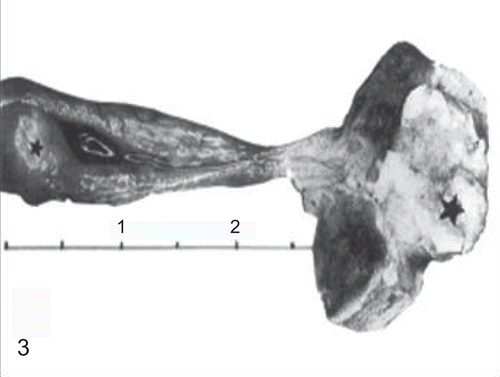
Figure 4. Opened duodenal lumen of a mouse treated with the high dose of folpet. Note the large nodule growing on the mucosa (arrow). (Reproduced by permission from A. Nyska et al., Induction of gastrointestinal tumors in mice fed the fungicide folpet: possible mechanisms. 1990. Jpn J Cancer Res 81:545–549.)
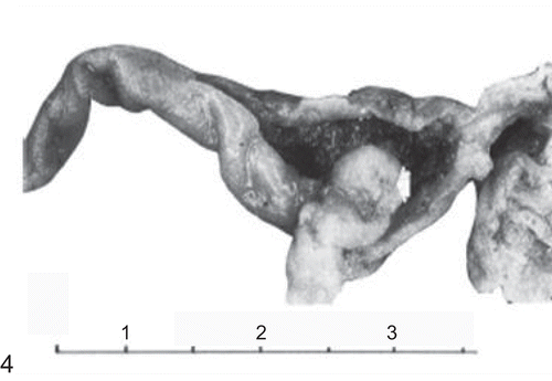
Figure 5. Histological section of the forestomach of a mouse treated with the high dose of folpet. Note squamous cell carcinoma infiltrating the wall (H&E, ×200). (Reproduced by permission from A. Nyska et al., Induction of gastrointestinal tumors in mice fed the fungicide folpet: possible mechanisms. 1990. Jpn J Cancer Res 81:545–549.)
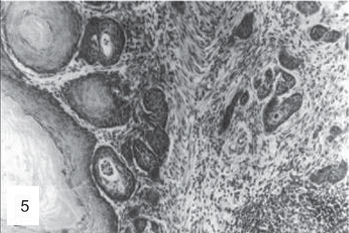
Figure 6. Histological section of a duodenal adenocarcinoma in a mouse treated with the high dose of folpet (H&E, ×200). (Reproduced by permission from A. Nyska et al., Induction of gastrointestinal tumors in mice fed the fungicide folpet: possible mechanisms. 1990. Jpn J Cancer Res 81:545–549.)
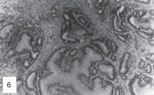
Table 3. Tumor incidences of selective tissues in a study in Sprague-Dawley rats administered folpet in the diet (60 rats per group) (CitationCox et al., 1985).
Table 4. Tumor incidences of selective tissues in a study in F344 rats administered folpet in the diet (20 rats per group) (CitationCrown et al., 1989).
Table 5. Tumor incidences of selective tissues in a study in F344 rats administered folpet in the diet (60 rats per group) (CitationCrown et al., 1985).
Table 6. Concordance table for analysis of potential human relevance of cytotoxicity mode of action of folpet induction of duodenal tumors in mice.
Figure 7. Diagrams illustrating the sequence of events in folpet-induced duodenal tumors. (A) Simplified diagram of normal duodenum showing normal crypt and villous structures. (B) Folpet-induced damage of duodenum with blunting of the villi and widening of the crypts. (C) Enlargement of crypts in response to duodenal damage, associated with increased proliferation and number of crypt cells. (D) Proliferation of crypt cells leading to development of adenoma, the precursor lesion for adenocarinoma.
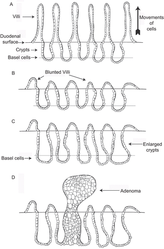
Table 7. Histopathologic changes in the forestomach of Sprague-Dawley rats administered folpet in the diet (CitationCox et al., 1985).