Figures & data
Figure 1. Schematic illustration of the chrysotile fiber. Chrysotile is a rolled sheet or concentrate rings of silicate with the magnesium on the outside of the sheet and the silica on the inside. The chrysotile fiber is acid soluble. Chrysotile has the formula Mg3Si2O5(OH)4. The fiber consists of magnesium hydroxide layers condensed onto silicon·oxygen tetrahedra. The fiber walls are made up of 11 to 21 such layers in which there is some mechanical interlocking. There is not any chemical bonding as such between the layers, however. Each layer is about 7.3 Å thick. The Mg(OH)2 part of the molecule layers is closest to the fiber surfaces; the silicon–oxygen tetrahedra are inside. Under the acid conditions associated with the macrophage, the fiber structure is weakened and the long fibers break into short pieces which can be engulfed and cleared by the macrophages.
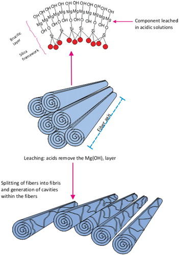
Figure 2. With amphiboles, the soluble cations shown as small circles are located between the fibers which are formed with double chain silicate. When the soluble cations dissolve as can happen in the lung, the amphibole fibers in these bundles are released as individual fibers. The double chain silicate amphibole fibers themselves are highly insoluble in both the lung fluids and in the macrophages.
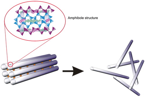
Table 1. Capabilities and limitations of analytical techniques used for asbestos measurements (reproduced from Berman & Crump, Citation2003)†.
Table 2. Epidemiological studies characterized as predominately chrysotile exposure by Hodgson & Darnton (Citation2000).
Table 3. Studies characterized as predominately chrysotile exposure (Hodgson & Darnton, Citation2000).
Figure 3. Map of North Charleston showing the location of the Textile plant (GARCO) and the US Navy Yard. The distance from GARCO to the Navy Yard is a few hundred meters. The width of the map is approximately 3.5 km.

Table 4. Concentrations of fiber and dust for workers in major sections of the Chongqin, China, asbestos plant, by job category, 1999. (Reproduced from Yano et al’s)*.
Table A1. Chronic inhalation studies with chrysotile.