Figures & data
Table 1. Formulation variables and dimensional properties of the microparticles.
Table 2. Calcium and GM content in microparticle samples.
Figure 1. SEM images of un-cross-linked (sample U) (a), cross-linked (sample A) (b), co-cross-linked (sample A1) (c), and co-cross-linked (sample A3) (d) microparticles.
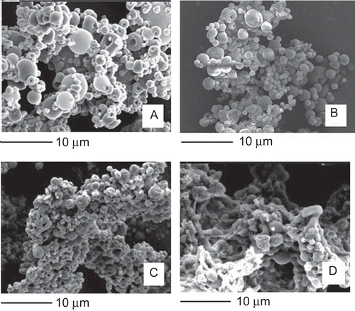
Figure 2. Size distribution of un-cross-linked (sample U), cross-linked (sample A), and co-cross-linked (sample A1) microparticles.
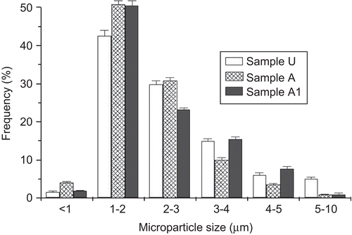
Figure 3. EDX analysis coupled with SEM: relative sodium and calcium percentages calculated on element emission intensity from distinct microparticles. The image refers to the sample A1.
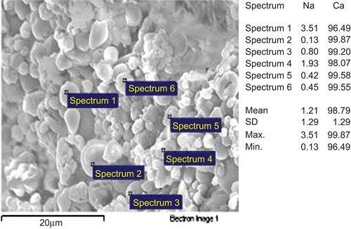
Figure 4. Antimicrobial activity of un-cross-linked (sample U), cross-linked (sample A), and co-cross-linked (sample A1) microparticles.
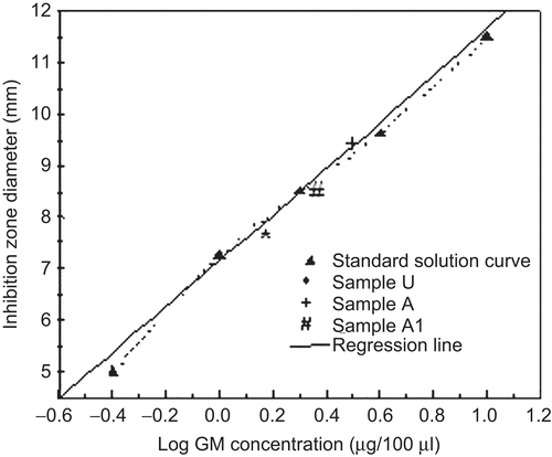
Figure 5. GM release from cross-linked microparticles in simulated gastrointestinal media.
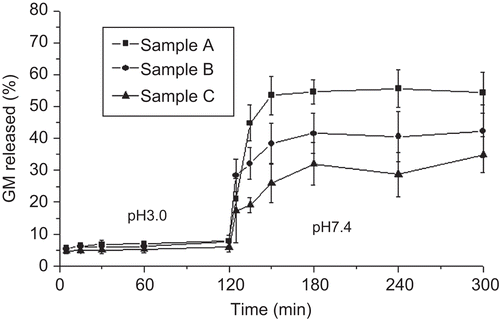
Figure 6. GM release from co-cross-linked microparticles compared with GM release from the sample A in simulated gastrointestinal media.
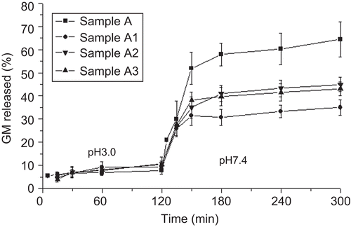
Figure 7. Epifluorescence microscopy image of the sample A1F recovered from simulated gastrointestinal media.
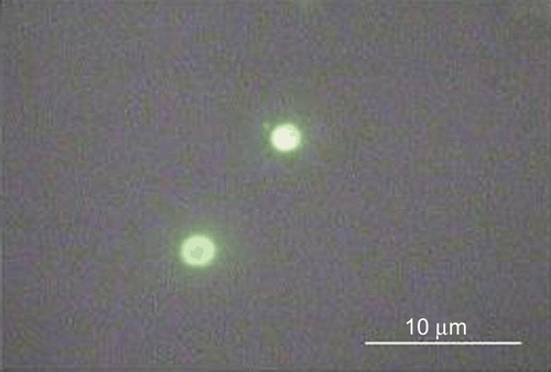
Figure 8. Image of a Peyer’s patch isolated from rat ileum.
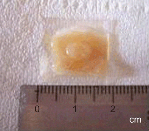
Figure 9. Interferential microscopy images of intestinal segments, including PP and perifollicular areas, from rabbit after perfusion treatment with microparticles: gut lumen surface (a), Bouin’s-fixed gut sections (spheroidal structures identified with microparticles are circled) (b). Original magnification ×40.
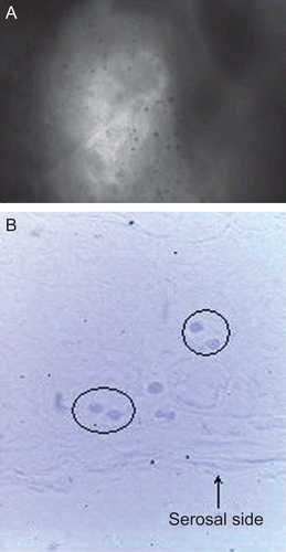
Figure 10. Fluorescence microscopy images of intestinal segments from rat after perfusion treatment with microparticles: gut lumen surface (a), perfused segment in correspondence of a PP (b), perfused segment in correspondence of intestinal villi (c). Original magnification ×40.
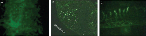
Figure 11. SEM image of the sample A1 after 30 min contact with pH 7.4 phosphate buffer solution.
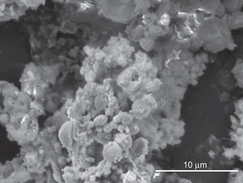
Figure 12. Cytofluorimetric analysis at 37°C (A) and 4°C (B): control (a), 3 h incubation (b), 6 h incubation (c).
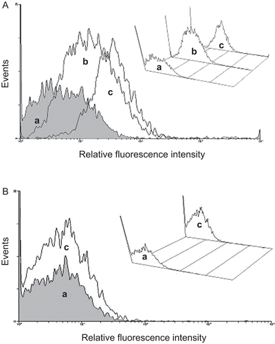
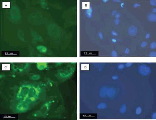
Figure 13. Confocal microscopy images of Caco-2 cells after nuclei staining: control under filter for green (a) and blue fluorescence (b); Caco-2 cells incubated at 37°C with the sample A1F under filter for green (c) and blue fluorescence (d).