Figures & data
Figure 1. Chemical structure of α-asarone.
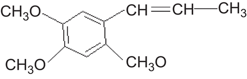
Figure 2. Schematic illustration of monolithic drug-in-adhesive patches.
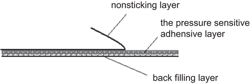
Table 1. Transdermal parameters of α-asarone patches through rat skin (x ± s, n = 3).
Figure 3. In vitro cumulative permeation amount per unit-time profiles of the transdermal patches containing α-asarone through rats skin. OA: oleic acid; IPM: Isopropyl myristate.
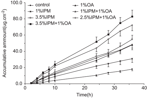
Table 2. Pharmacokinetics parameters of α-asarone patches in rabbits (n = 5).
Figure 4. Plasma concentration-time profile obtained after transdermal administration of α-asarone drug-in-adhesive patch on rabbits. Each time point represents mean ± SD (n = 5).
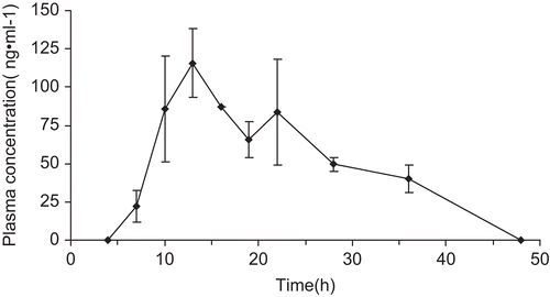
Figure 5. The number of inflammatory cells in BALF in different groups. BALF cells were separated using Cytospin and then stained with Wright. Differential cell counting was performed using standard morphological criteria. This experiment used six rats per group (n = 6). * p < 0.05, ** p < 0.01, *** p < 0.001 vs model; # p < 0.05, ## p < 0.01, ### p < 0.001 vs control. Total cells: the total cell number; EOS%: Eosinophil number; DXM: dexamethasone; Low dose: 4 cm2 of DIA; High dose: 1.5 cm2 of DIA; DIA: drug-in-adhesive.
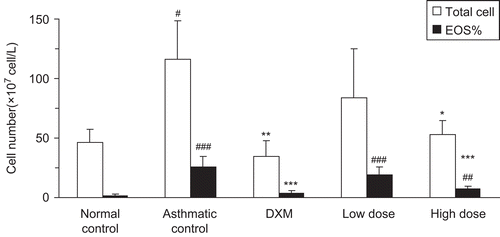
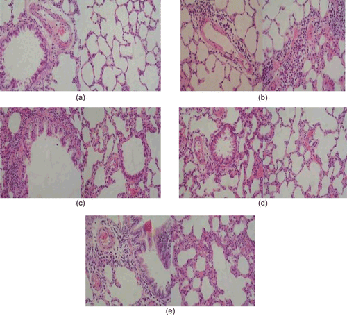
Figure 6. The cut sheets of the lung. Sections were stained by hematoxylin and eosin (200×): (a) normal control rat; (b) asthmatic control group; (c) treated with DXM; (d) treated with high dose of α-asarone (4 cm2); (e) treated with low dose of α-asarone (1.5 cm2).