4,086
Views
133
CrossRef citations to date
0
Altmetric
Original Article

Thymoquinone in liposomes: a study of loading efficiency and biological activity towards breast cancer

, , , &
Pages 371-377 | Received 20 Jun 2012, Accepted 03 Sep 2012, Published online: 08 Oct 2012

Figures & data

Figure 1.  Structure of thymoquinone (TQ).

Figure 1.  Structure of thymoquinone (TQ).

Figure 2.  Electron microscopic images for extruded TQ encapsulated liposomes; (A) SEM image (B) TEM image. The average diameter of liposomes is about 100 nm.
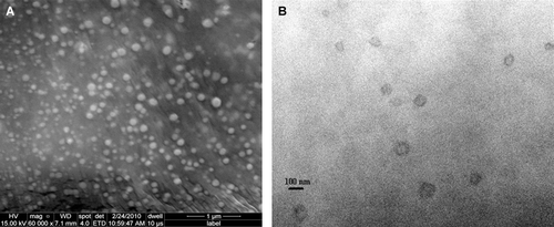
Figure 2.  Electron microscopic images for extruded TQ encapsulated liposomes; (A) SEM image (B) TEM image. The average diameter of liposomes is about 100 nm.

Figure 3.  Optical microscope image of non-extruded TQ-LP.

Figure 3.  Optical microscope image of non-extruded TQ-LP.

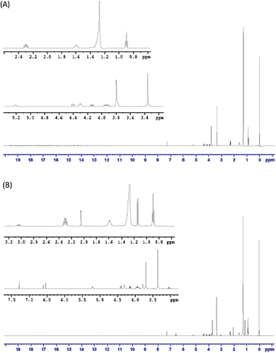
Figure 4.  1H-NMR spectrum for A) DPPC and B) DPPC + TQ mixture in CDCl3.

Figure 4.  1H-NMR spectrum for A) DPPC and B) DPPC + TQ mixture in CDCl3.

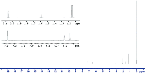
Figure 5.  1H-NMR spectrum for TQ in CDCl3.

Figure 5.  1H-NMR spectrum for TQ in CDCl3.

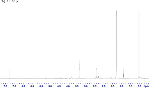
Figure 6.  1H-NMR spectrum for TQ-LP in CDCl3.

Figure 6.  1H-NMR spectrum for TQ-LP in CDCl3.

Figure 7.  Differential scanning calorimetry (DSC) analysis. The melting temperatures (Tm) (sharp peaks) of liposomes are compared. The Tm of DPPC liposome (41.8°C) was decreased by 2°C compared with DPPC-Triton X100 liposome (39.8°C).
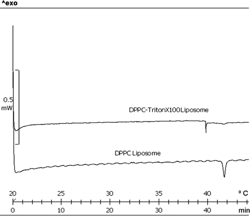
Figure 7.  Differential scanning calorimetry (DSC) analysis. The melting temperatures (Tm) (sharp peaks) of liposomes are compared. The Tm of DPPC liposome (41.8°C) was decreased by 2°C compared with DPPC-Triton X100 liposome (39.8°C).

Figure 8.  Anti-proliferative effects of liposomal TQ on cell lines MCF-7 (A) T47D (B) and PLF (C). Readings are the mean of two replicate experiments each was done in triplicate.
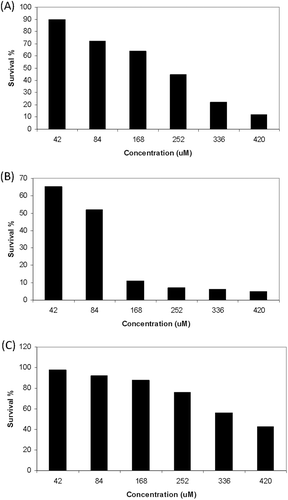
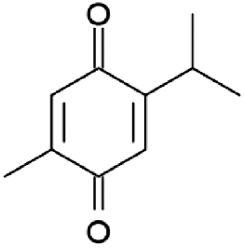
Figure 8.  Anti-proliferative effects of liposomal TQ on cell lines MCF-7 (A) T47D (B) and PLF (C). Readings are the mean of two replicate experiments each was done in triplicate.
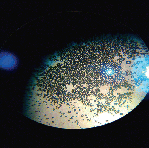
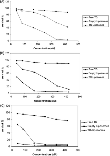
Figure 9.  Comparison of cytotoxicity between liposomal TQ, free TQ and empty liposomes on periodontal ligament fibroblasts cells (a), MCF-7 (b) and T47D (c).

Figure 9.  Comparison of cytotoxicity between liposomal TQ, free TQ and empty liposomes on periodontal ligament fibroblasts cells (a), MCF-7 (b) and T47D (c).

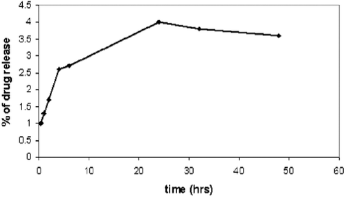
Figure 10.  The in vitro release of thymoquinone from TQ-LP liposomes.

Figure 10.  The in vitro release of thymoquinone from TQ-LP liposomes.

Reprints and Corporate Permissions

Please note: Selecting permissions does not provide access to the full text of the article, please see our help page How do I view content?

To request a reprint or corporate permissions for this article, please click on the relevant link below:

Academic Permissions

Please note: Selecting permissions does not provide access to the full text of the article, please see our help page How do I view content?

Obtain permissions instantly via Rightslink by clicking on the button below:

If you are unable to obtain permissions via Rightslink, please complete and submit this Permissions form. For more information, please visit our Permissions help page.