
Figures & data
Figure 1. Locations of entrapped hydrophilic, lipophilic and amphipathic drugs in niosomes.
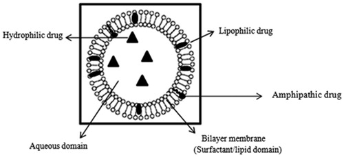
Figure 2. Major types of niosomes, MLV (multilamellar vesicles), OLV (oligolamellar vesicles) and ULV (unilamellar vesicles). Small circles (o) represent polar head group, sticks (–) represents apolar tails of single-chain surfactant molecules and a bilayer membrane represent a circular double surfactant molecules layer oriented in continuous tails-to-tails and polar heads lining the inner and outer circle.
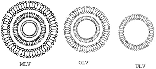
Figure 3. Schematic of a single-chain surfactant, ao = hydrophilic head group area, v = hydrophobic chain volume, lc = hydrophobic chain length.
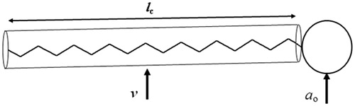
Table 1. Geometrical characteristic of surfactants and proposed various surfactant aggregates. Bulky head groups’ surfactants form micelles, cone-shaped surfactants form inverted micelles and cylindrical-shaped surfactants form bilayers.
Figure 4. Chemical structure of cholesterol.
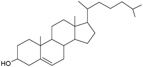
Figure 5. Hypothetical positions occupied by DCP (A), Solulan (B) and sodium cholate (C) in the bilayer membrane of Span 60 niosomes stabilised by cholesterol (Abdelkader et al., 2011).
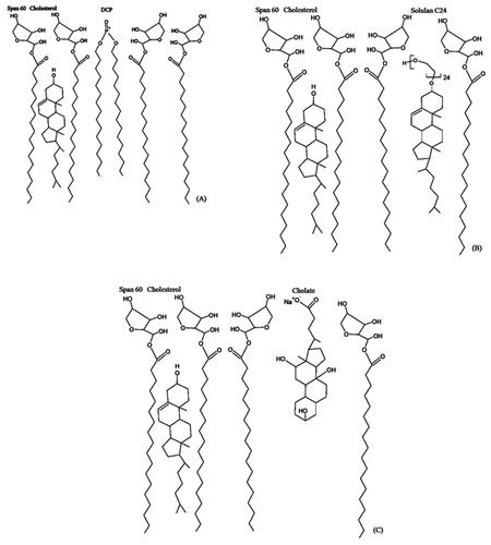
Table 2. Chemical structure, molecular weight, CMC and phase transition temperature of ionic and surfactant additives.
Figure 6. Confocal laser scanning micrographs of discomes (left) and conventional spherical niosomes (right) loaded with carboxyfluorescein (Abdelkader et al., 2012b).
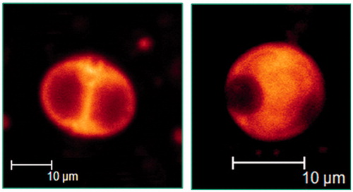
Table 3. Overview of main characterization techniques for niosomes.