Figures & data
Table 1. Types of prepared ethambutol hydrochloride niosomal formulations with mean percent entrapment.
Table 2. ZP, mean particle diameters and PI of investigated niosomes.
Figure 3. Mean percent ethambutol hydrochloride released from F2 and F4 in phosphate buffer saline (pH 7.4).
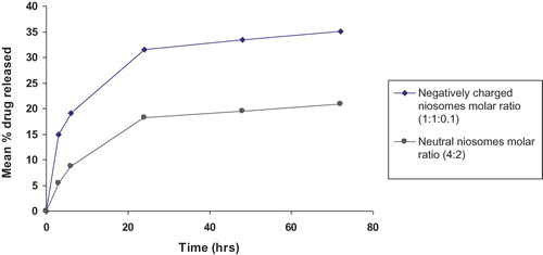
Figure 4. Mean amount of ethambutol hydrochloride deposited in mice lungs (ng/mg lung) after single s.c. injection of free drug compared with F2 and F4.
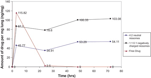
Figure 5. AUC of ethambutol HCl deposited in mice lungs (ng/mg lung) after single s.c. injection of the three treatment groups.
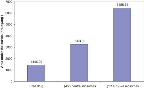
Table 3. Mean RSLW values for lungs and bacterial counts of guinea pig lung homogenates receiving different treatments.
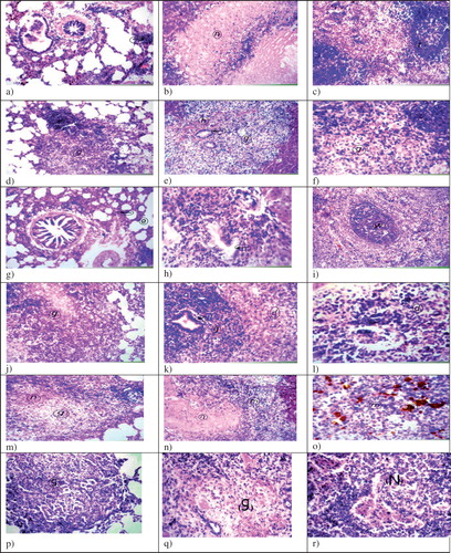
Figure 6. Histopathological examination of guinea pigs organs infected and differently treated (H&E, ×40), (a) Lung infected and treated with free ethambutol hydrochloride 6 days weekly for 6 weeks, show focal lymphoid cells aggregation (arrow) and diffuse lymphoid cells infiltration in between the air alveoli, (b) Livers show wide circumscribed area of necrosis replacing the hepatic parenchyma (n), (c) Spleens show mild depletion in central portion of the white pulp (f), (d) Lungs infected and treated with free ethambutol hydrochloride twice weekly for 6 weeks, show granuloma formation mainly cellular (g) with central necrosis, (e) Livers show granuloma formation (g) with haemorrhages (h) and hyperplasia in bile duct (arrow), (f) Spleens show the necrosis in the lymphoid cells in the center of the white pulps (g), (g) Lungs infected and treated with ethambutol hydrochloride niosomes (1:1:0.1 negative) (F2) twice weekly for 6 weeks, show collapse (arrow) and emphysema (e) in air alveoli, (h) Livers show hyperplasia in bile duct (arrow) in the portal area, (i) Spleens show intact white pulp (w) with hemosiderosis in red pulp, (j) Lungs infected and treated with ethambutol hydrochloride niosomes (4:2 neutral) (F4) twice weekly for 6 weeks, show cellular granuloma with central necrosis (g) replacing the lung parenchyma, (k) Livers show granuloma formation mainly from lymphoid cells (g) with hyperplasia in bile duct (arrow) and degeneration in other hepatocytes (d), (l) Spleens show lymphoid cellular necrosis in the white pulp (d), (m) Lungs of the control group infected and receiving saline, show granuloma with central necrosis replacing the lung parenchyma (g, n), (n) Livers show necrosis in the central zone of the granuloma (n) surrounded by other leucocytes (m), (o) Spleens show diffuse proliferation of epitheloid cells (m) with necrobiosis in the lymphoid cells and diffuse hemosiderosis (h), (p) Lungs of the control group infected and receiving neutral drug-free niosomes of the molar ratio Span 60: cholesterol (4:2), show cellular granuloma formation without central necrosis (g), (q) Livers show granuloma formation with central necrosis without fibrous connective tissue capsule formation (g) and (r) Spleens show cellular granuloma formation with central necrosis (N).