Figures & data
Table 1. Characterization of CPT NPs.
Figure 1. (A) Size distribution of CPT-loaded biotin-F127-PLA NPs and (B) transmission electron micrograph of the targeted CPT NPs.
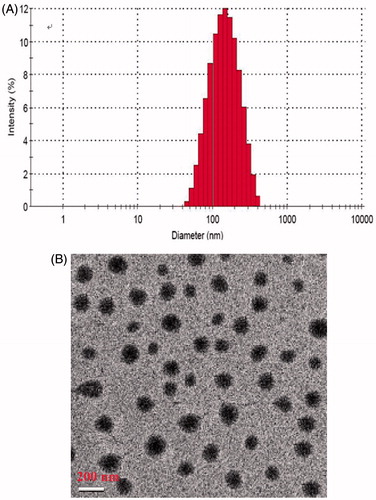
Table 2. Evaluation of antibody binding rate and coupling ratio of antibody to NPs.
Figure 2. In vitro release profile of CPT from NPs in phosphate buffer solutions.
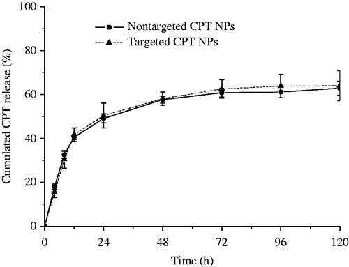
Figure 3. In vitro antitumor effects of CPT NPs on H22 cell lines. The concentrations of the blank NPs (control group) were set at the same level as those in the drug-loaded NPs group. **p < 0.01 compared with the control group.
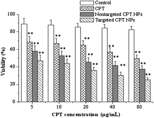
Figure 4. Tissue distribution of CPT in tumor-bearing mice after intravenous administration of non-targeted NPs (A) and targeted NPs (B). *p < 0.05 compared with the plasma.
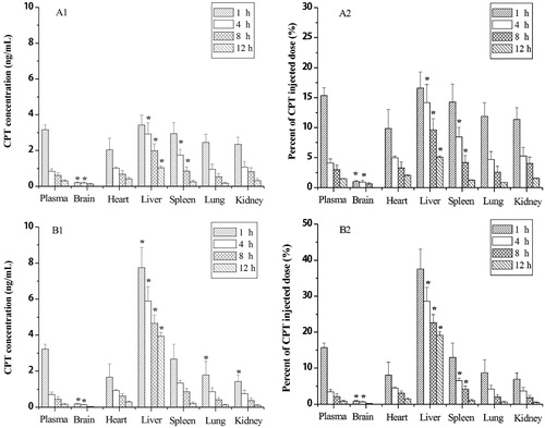
Figure 5. In vivo antitumor effect of CPT in either targeted or non-targeted CPT NPs in H22-bearing mice. Tumor weight (A) and tumor inhibition rate (B). *p < 0.05 compared to the free CPT group.
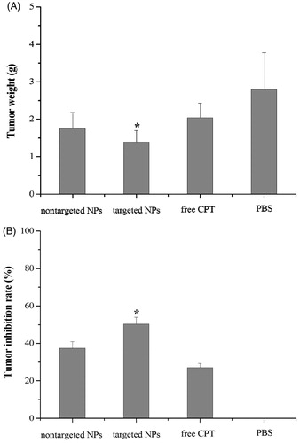
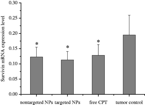
Figure 6. Survivin mRNA expression of mice bearing H22 tumor tissue. *p < 0.05 compared with the control group.