Figures & data
Figure 1. Digital photographs of C57BL/6 mouse dorsal skin with and without treatment with the Vaniqa eflornithine cream (13.9%) for up to 36 days. The hair on the application area was removed by plucking using GiGi® Honee warm wax, chemical depilation using Nair® or trimming with a clipper. In one group (bottom), following trimming, the skin area was also treated with a microneedle roller (microneedle length, 500 µm; base diameter, 50 µm) every time before the application of the eflornithine cream. The rectangles indicate the mouse skin area where the eflornithine cream was applied. For a full-color version of the figure, the reader is referred to the online version of the article.
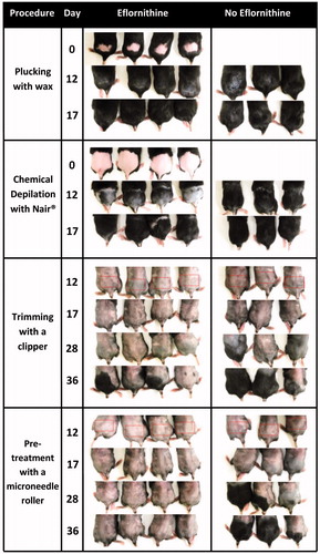
Figure 2. In vitro permeation of eflornithine hydrochloride in a solution through a mouse skin area where the hair was trimmed (without microneedle) or trimmed and then treated with microneedles (with microneedle). Data shown are mean ± SD (n = 3).
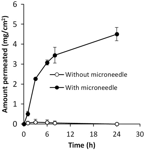
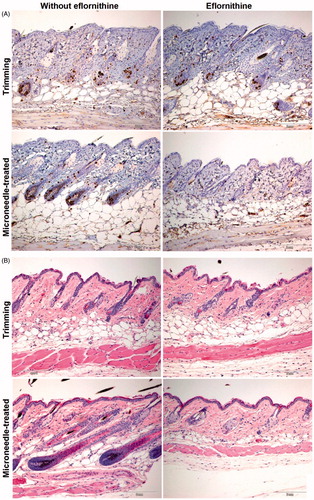
Figure 3. Representative micrographic pictures of skin samples after anti-BrdU staining (A) or H&E staining (B). Scale bar = 2 mm. For a full-color version of the figure, the reader is referred to the online version of the article.