Figures & data
Table 1. Effect of the phospholipid/cholestenone ratio on physicochemical properties of DOX-loaded liposomes.
Figure 1. Zeta potential of blank liposomes (A), SA-added liposomes (B), and HA-modified liposomes (C).
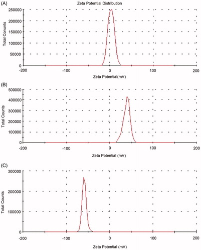
Figure 2. TEM morphology of DOX-loaded liposome.
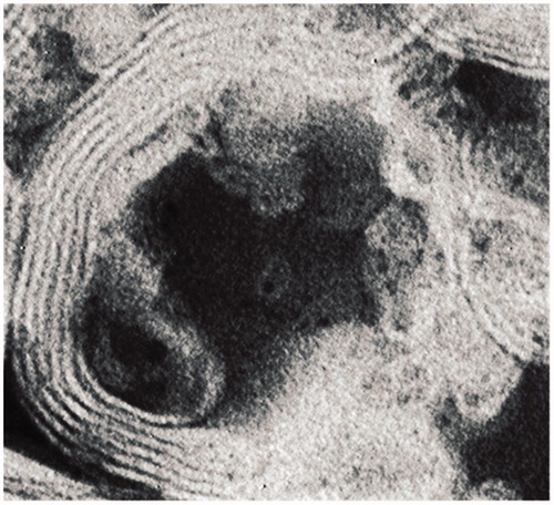
Figure 3. In vitro release profile of DOX solution and liposomes in PBS solution (pH = 7.4) at 37 °C (n = 3, mean ± SD).
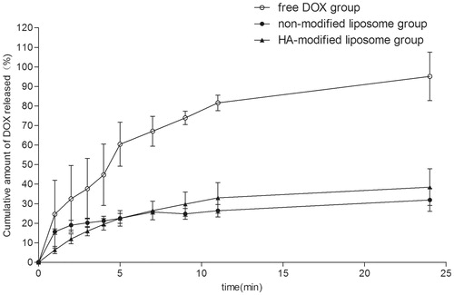
Figure 4. Cytotoxicity of HA-modified liposomes in human epithilium cells.
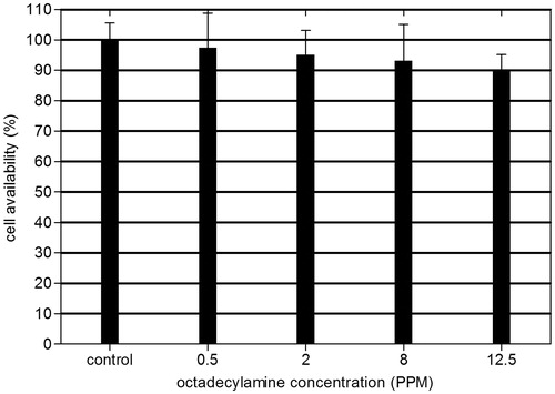
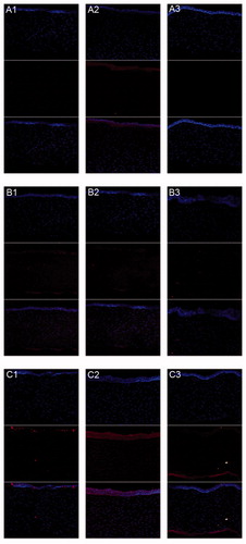
Figure 5. CLSM of DOX uptakes by human corneal epithelia cell after incubated alternately with 40 PPM (A1–A3) free DOX group, (B1–B3) non-modified liposome group, and (C1–C3) HA-modified liposome group after (A1, B1, and C1) 0.5 h, (A2, B2, and C2) 3 h, and (A3, B3, and C3) 6 h. (Up) blue filter; (middle) red filter; (down) overlay of both (up and middle) sections.
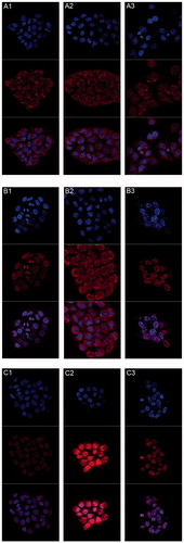
Figure 6. In vitro transcorneal permeation of DOX liposome preparation and solution (X ± SD, n = 3).
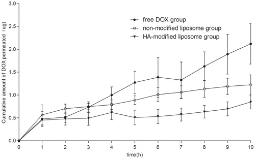
Figure 7. Kinetics of DOX disappearance from tear fluid following topical administration of the liposome and the control solution (X ± SD, n = 4).
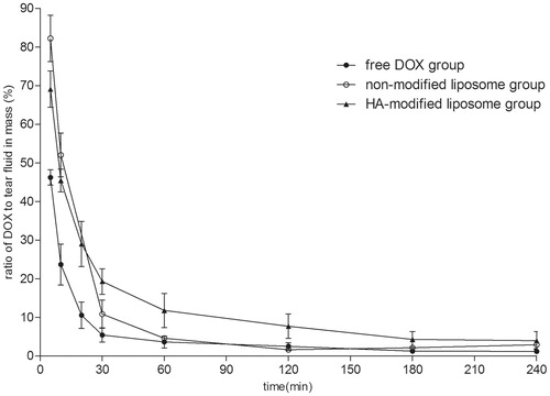
Figure 8. Concentration–time profles of DOX in aqueous humor after instillation of 0.5 mg/mL DOX liposome preparation and DOX solution in rabbits (ng/mL, X ± SD, n = 4).
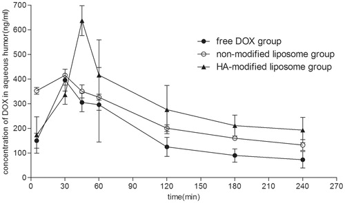
Table 2. Pharmacokinetic parameters of DOX in the aqueous humor of rabbits (n = 4).
Figure 9. CLSM of DOX absorption by corneal tissues after rabbits’ eyes in vivo treated alternately with free DOX (A1–A3), (B1–B3) non-modified liposome, and (C1–C3) HA-modified liposome after (A1, B1, and C1) 0.5 h, (A2, B2, and C2) 2 h, and (A3, B3, and C3) 3 h. (Up) blue filter; (middle) red filter; (down) overlay of both (up and middle) sections.