Figures & data
Figure 1. Nano-hydroxyapatite artificial bone under inverted microscope.
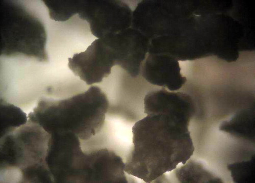
Figure 2. Nano-hydroxyapatite artificial bone under scanning electron microscope.
Table I. MTT analysis of bone marrow mesenchymal stem cell proliferation (_x ± s, n = 12, A value).
Figure 3. Primary cells covered the bottom of culture flask, orderly arranged along long axis of cells body.
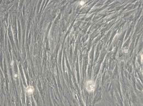
Figure 4. Osteogenic induction for 21 d, alizarin red staining of calcium nodules were red.
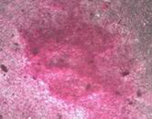
Figure 5. Cells transfected by adenovirus-green fluorescent protein 48 hour were almost green fluorescent.
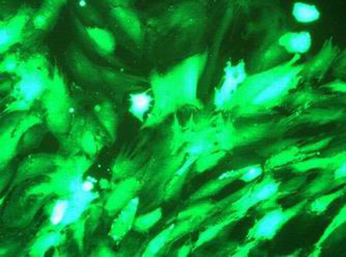
Figure 6. Transfection 72 hour, bone morphogenetic protein-2 immunohistochemical staining showed positive expression in cytoplasm.
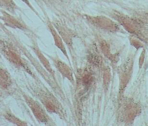
Figure 7. Untransfected cells were negative for bone morphogenetic protein-2 immunohistochemical staining.
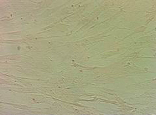
Figure 8. Western blot detection of bone morphogenetic protein-2 expression.
Figure 9. Compound for 3 d, transfected bone marrow mesenchymal stem cells adhere and extend good in the lacunae and loophole of nano-hydroxyapatite material.
Figure 10. Compound for 5 d, transfected bone marrow mesenchymal stem cells fully spread and extend pseudopodia on the surface of nano-hydroxyapatite A material, calcium deposit can be seen around the cells.
Figure 11. Western blot detection of bone morphogenetic protein-2 expression in cells.