Figures & data
Figure 1. The EarMark™ system as arranged for a patient's radiographic studies (i.e., CT scanning). At top left is a side view, with an overhead view at top right. The EarMark™ is composed of carbon fiber. It is coupled to the LADS (which has the appearance of a mouthguard) via a customized block epoxied to the EarMark™, allowing a screw-fit to the LADS. At bottom is the numbering scheme for the fiducial markers. These numbers are used in data reporting in , , and .
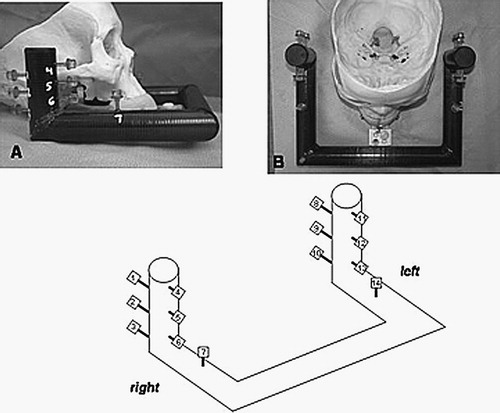
Figure 2. The EarMark™ system as arranged for operative use. Panel A shows a side view, Panel B an overhead view, and Panel C a frontal view. The mouthpiece-like LADS is seen in blue with a Plexiglas extender jutting out anteriorly. The infrared (IR) emitter is the black X-shaped device. This is connected to the LADS via the customized adapter (in white) which allows positioning of the IR emitter in an identical fashion both with and without the EarMark™.
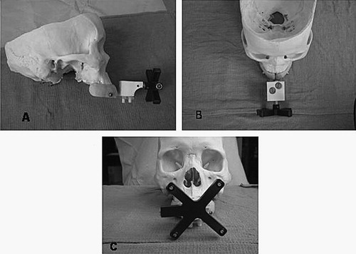
Figure 3. The experimental set-up for determining target registration error within the region of the temporal bone. Illustrated is one of the three skulls which has had the temporal bones removed and replaced with 2 discs, each holding 13 surgical targets arranged in a cross-hair pattern over the approximate centroid of the temporal bone. The left photograph shows the vertical disc while the right photograph shows the horizontal disc. The respective identification systems are shown in the schematics beneath the photographs. These identifiers are used in data reporting in , and .
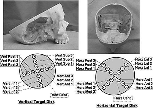
Figure 4. Graphical representation of Target Registration Errors (TREs). Shown are 3D reconstructions of the skull and target system from the CT scans. The surgical targets are colored according to error (blue=least error, red=most error; a numerical scale corresponding to colors is shown at right). The left panel presents the horizontal disc best, while the right panel presents the vertical disc best.
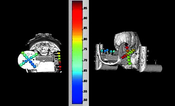
Figure 5. Graphical representation of the Fiducial Registration Errors (FREs). Shown are 3D reconstructions of the skull and fiducial frame from the CT scans. The fiducials are colored according to error (blue=least error, red=most error; a numerical scale corresponding to colors is shown at right). The left panel presents the left side (fiducials #8–14), while the right panel presents the right side (fiducials #1–7) (see for fiducial numbering scheme).
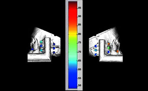
Table I. Target registration errors for Skull 1.
Table II. Fiducial registration errors for Skull 1.
Table III. Target registration errors for Skull 2.
Table IV. Fiducial registration errors for Skull 2.
Table V. Target registration errors for Skull 3.
Table VI. Fiducial registration errors for Skull 3.