Figures & data
Figure 1. Post-operative radiograph of total hip arthroplasty with a U2 Total Hip Joint (United Orthopedic Corporation, Hsinchu, Taiwan). We marked the outer shell of the acetabulum with an ellipse (s = the short axis of the ellipse; l = the long axis). Wan et al. described the measured angle α as acetabular radiographic version Citation[15]. Because of the symmetry of the ellipse, angle α is equal to angles β, γ, and δ. We previously used a special protractor to measure angle β for acetabular radiographic version Citation[7].
![Figure 1. Post-operative radiograph of total hip arthroplasty with a U2 Total Hip Joint (United Orthopedic Corporation, Hsinchu, Taiwan). We marked the outer shell of the acetabulum with an ellipse (s = the short axis of the ellipse; l = the long axis). Wan et al. described the measured angle α as acetabular radiographic version Citation[15]. Because of the symmetry of the ellipse, angle α is equal to angles β, γ, and δ. We previously used a special protractor to measure angle β for acetabular radiographic version Citation[7].](/cms/asset/503423c1-9075-43d4-b1c0-d1f97d4a2dfa/icsu_a_583805_f0001_b.gif)
Figure 2. (a) Theoretical error of Wan's method. (b) Error correction chart for Wan's method. After obtaining a measurement using Wan's method, the surgeon can add the correction angle from this chart.
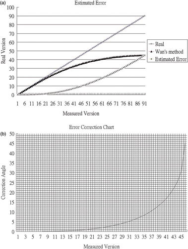
Figure 3. Error with Wan's method before and after mathematical correction.
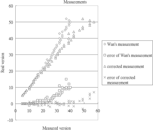
Table I. Results obtained with simulated radiographs.
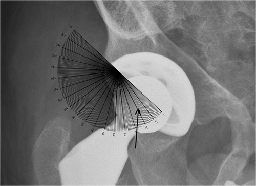
Figure 4. We use an ordinary goniometer to measure acetabular radiographic version directly. In this example, the goniometer is aligned with the long axis of the ellipse (the metal ring) and Wan's angle is then read on the middle point of the ellipse (arrow).