Figures & data
Table I. Patient data.
Figure 1. Stages of the gender-specific pelvis SSM generation pipeline. The SSM generation procedure consists of the following consecutive steps: segmentation of pelvic bone structures from the collected CT datasets; generation of the male and female training sets; mesh alignment based on Procrustes analysis; and, finally, application of principal component analysis (PCA) to obtain the gender-specific SSM.
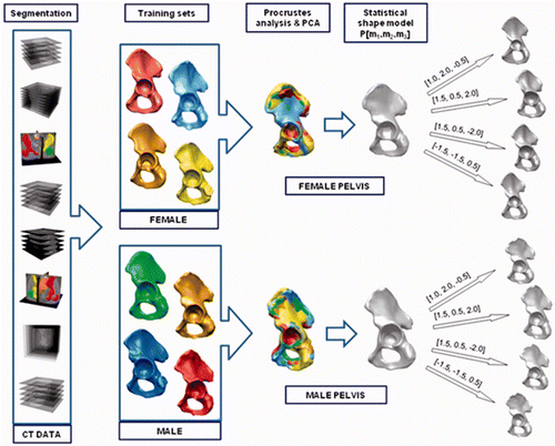
Table II. Volumetric characterization of the hemipelvises in the gender-specific pelvic SSMs.
Table III. Statistical view of the distance between the anterior superior iliac spine (ASIS) and the center of the sphere inscribed in the acetabulum for all pairs of hemipelvises (male and female) in both training sets.
Table IV. Percentage of hemipelvis surface to be reconstructed.
Figure 2. Application and evaluation of the SSM-based pelvic reconstruction approach in clinical cases. First column: Location of the defects (tumors) in the segmented CT data, marked with red circles. Second column: Reconstructed hemipelvis surface (shown in yellow). Third column: Color-coded map of the distances between the original and the reconstructed pelvic surfaces. The uncolored portion is the reconstructed defect area (after virtual wide resection of the bone tumor). Fourth column: Distribution of the distances between the original and the reconstructed (SSM-based) pelvic surfaces in the intact areas, and the average distance (red dotted line). Fifth column: Map of distances between the original and the mirroring-based reconstructed pelvic surfaces (in intact areas). Sixth column: Distribution of the distances for the reconstruction method in the preceding column. Seventh column: Distance maps for the SSM-based reconstruction of the equivalent osteotomy performed in the contralateral healthy hemipelvis (for the whole hemipelvis). The contralateral healthy hemipelvis acts as the ground truth in this part of the evaluation. Eighth column: Distribution of the distances for the reconstruction method in the preceding column.
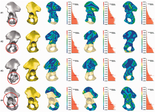
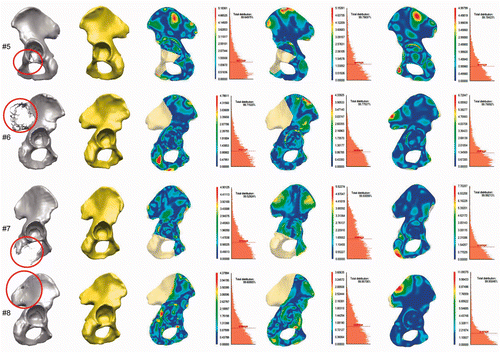
Table V. Mean distances from original pelvis to SSM-based reconstruction and mirroring method-based reconstruction for all evaluated cases (intact area only).
Table VI. Mean distance differences for SSM-based reconstruction and mirroring method-based reconstruction for each case and degree of improvement.
Table VII. Mean distances from contralateral healthy hemipelvis to SSM-based reconstruction of equivalent osteotomy performed in contralateral healthy hemipelvis (presented separately for intact area only, defect area only, and whole hemipelvis area).
Figure 3. Convergence behavior of the objective function during the evolution strategy-based optimization process for each reconstruction case. Equilibrium is achieved after 80 generations, on average.
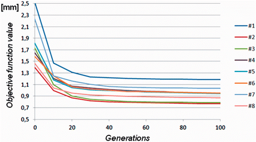
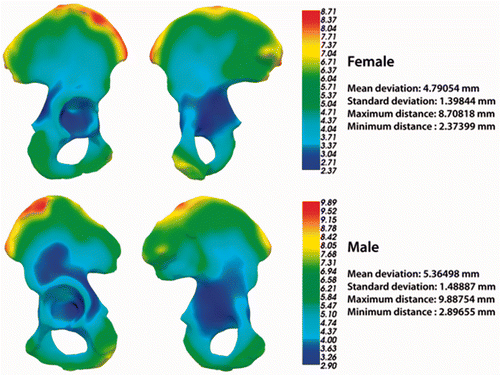
Figure 4. Pelvis shape variability maps based on the male and female pelvic surfaces from both training sets.