Figures & data
Figure 1. Selected histological findings: (A) and (B) hematoxylin-eosin stains. (A) photomicrograph showing normal lining thickness with increased stromal cellularity in part due to infiltrating large mononuclear cells. The arrow points to large macrophages within focal amyloid deposits (patient 1). (B) Example of mild focal lining hyperplasia and juxta-intimal pericapillaritis occasionally seen in the specimen from patient 2. (C) and (D) Detection of amyloid by Congo Red stain (patient 1). (C) Typical carmine red deposits as seen in bright field view. (D) Typical birefringence seen under polarizing light. (E–G) Immunohistochemical detection of kappa light chain (patient 1). There is diffuse deposition of kappa (E), but not of lambda (F) light chains, consistent with AL amyloidosis. G, negative control.
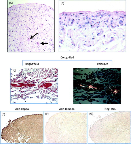
Figure 2. Immunohistochemical findings. Immunohistochemical stains for CD68, CD3, CD20 and CD38. Chromogen, DAB (brown). Original magnification, 100×. Left panel: synovial biopsy from patient 1. Middle panel: paraffin-embedded synovial fluid sediment from patient 1. Right panel: synovial biopsy from a patient with RA with active disease despite treatment with disease-modifying antirheumatic drugs.
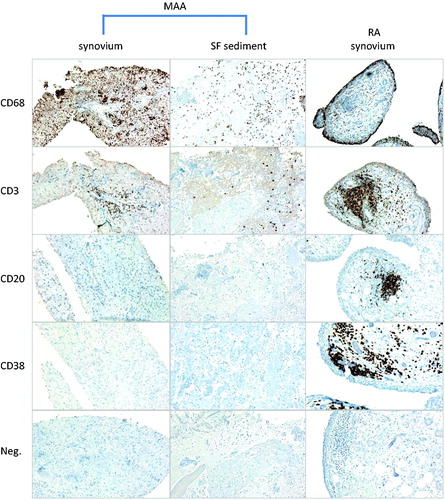
Figure 3. Cluster analysis of the 2 MAA specimens with respect to RA, OA and normal synovium. Expression values in the synovial subintima (number of positive staining cells per mm2) of CD68, CD3, CD20, CD38, Ki-67 and vWF for MAA (n = 2), RA (n = 24), OA (n = 26) and normal synovium (n = 15) were used to construct a hierarchical tree of clusters for all specimens. The MAA specimens fall within a cluster of OA specimens and are clearly separated from the RA specimens.
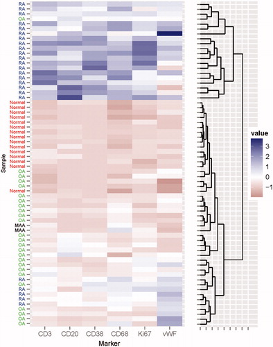
Table 1. Demographic and laboratory data.
Table 2. Summary of histomorphometric results.
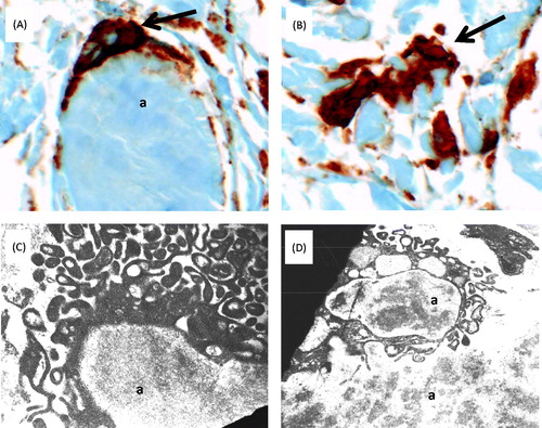
Figure 4. Amyloid phagocytosis in synovial tissue from patient 1. (A) and (B) Immunohistochemial detection of CD68+ macrophages (brown) engulfing (A) and interdigitating with (B) amorphous amyloid deposits (original magnification 400×). (C) and (D) Ultrastructural findings. (C) Phagocyte reacting to and surrounding a mass of amyloid (a); magnification approximately 12 000×. (D) Cell with completed amyloid phagocytosis; magnification approximately 7000×. Images were acquired with a Zeiss EM-10 transmission electron microscope, using thin sections from epoxy-embedded tissue. Tissue was obtained during the same biopsies as in . Abbreviation: a, amyloid.