10,712
Views
119
CrossRef citations to date
0
Altmetric
Review Article

Biomarkers of kidney injury

, &
Pages S22-S30 | Received 13 Dec 2010, Accepted 07 May 2011, Published online: 28 Jun 2011

Figures & data

Figure 1.  Inner cortex of a rat kidney shown after 24 h of reperfusion following experimental bilateral renal ischemia by clamping of both renal pedicles for 60 min. Kidneys were perfusion-fixed with paraformaldehyde and stained with hematoxylin and eosin (H&E). Slightly dilated tubules of the S3-segments of the proximal tubule are packed with detached cells in the lumen (examples are indicated by arrows), the basal membrane is denuded. Congested vasa recta (in middle) are filled blood cells (from N. Obermüller, personal data).
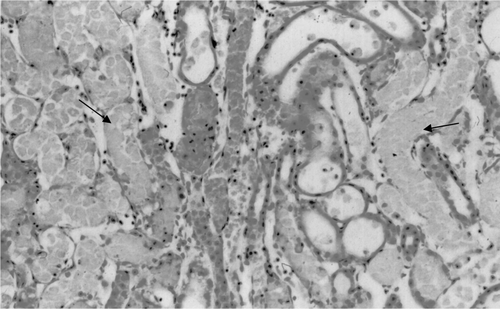
Figure 1.  Inner cortex of a rat kidney shown after 24 h of reperfusion following experimental bilateral renal ischemia by clamping of both renal pedicles for 60 min. Kidneys were perfusion-fixed with paraformaldehyde and stained with hematoxylin and eosin (H&E). Slightly dilated tubules of the S3-segments of the proximal tubule are packed with detached cells in the lumen (examples are indicated by arrows), the basal membrane is denuded. Congested vasa recta (in middle) are filled blood cells (from N. Obermüller, personal data).

Table 1.  Characteristics of AKI biomarkers.

Reprints and Corporate Permissions

Please note: Selecting permissions does not provide access to the full text of the article, please see our help page How do I view content?

To request a reprint or corporate permissions for this article, please click on the relevant link below:

Academic Permissions

Please note: Selecting permissions does not provide access to the full text of the article, please see our help page How do I view content?

Obtain permissions instantly via Rightslink by clicking on the button below:

If you are unable to obtain permissions via Rightslink, please complete and submit this Permissions form. For more information, please visit our Permissions help page.