345
Views
5
CrossRef citations to date
0
Altmetric
Research Article

Diazoxide supplemented Celsior solution improves hypothermic heart preservation effect in rat through activation of mitochondrial ATP-sensitive potassium channel

, , , , &
Pages 1060-1066 | Received 03 Jul 2008, Accepted 26 Aug 2008, Published online: 19 Oct 2009

Figures & data

Figure 1. Time changes of left ventricular developed pressure (LVDP) (% of equilibrium values) during 60 min of reperfusion after hypothermic preservation. (A) 3 h of preservation; (B) 8 h of preservation. Data are expressed as mean ± SD, n = 8. *p < 0.05, **p < 0.01 vs. control group; #p < 0.05, ##p < 0.01 vs. 30 μM DE group; +p < 0.05, ++p < 0.01 vs. 15 μM DE group.
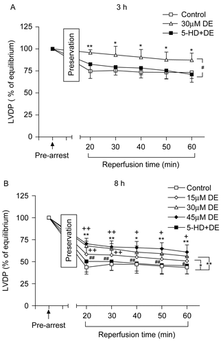
Figure 1.  Time changes of left ventricular developed pressure (LVDP) (% of equilibrium values) during 60 min of reperfusion after hypothermic preservation. (A) 3 h of preservation; (B) 8 h of preservation. Data are expressed as mean ± SD, n = 8. *p < 0.05, **p < 0.01 vs. control group; #p < 0.05, ##p < 0.01 vs. 30 μM DE group; +p < 0.05, ++p < 0.01 vs. 15 μM DE group.

Figure 2. Recovery of coronary flow (CF) (% of equilibrium values) at the end of reperfusion after hypothermic preservation. Data are expressed as mean ± SD, n = 8. *p < 0.05, **p < 0.01 vs. control group; #p < 0.05, +p < 0.05 vs. 15 μM DE group.
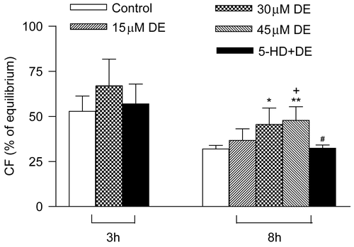
Figure 2.  Recovery of coronary flow (CF) (% of equilibrium values) at the end of reperfusion after hypothermic preservation. Data are expressed as mean ± SD, n = 8. *p < 0.05, **p < 0.01 vs. control group; #p < 0.05, +p < 0.05 vs. 15 μM DE group.

Figure 3. Release of myocardial lactate dehydrogenase (LDH) in coronary effluent before arrest and during reperfusion after hypothermic preservation. (A) 3 h of preservation; (B) 8 h of preservation. Data are expressed as mean ± SD, n = 8. *p < 0.05, **p < 0.01 vs. control group; ##p < 0.01 vs. 30 μM DE group.
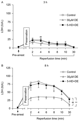
Figure 3.  Release of myocardial lactate dehydrogenase (LDH) in coronary effluent before arrest and during reperfusion after hypothermic preservation. (A) 3 h of preservation; (B) 8 h of preservation. Data are expressed as mean ± SD, n = 8. *p < 0.05, **p < 0.01 vs. control group; ##p < 0.01 vs. 30 μM DE group.

Figure 4. Release of myocardial creatine kinase (CK) in coronary effluent before arrest and at the 4th min of reperfusion after hypothermic preservation. Data are expressed as mean ± SD, n = 8. *p < 0.05, **p < 0.01 vs. control group; #p < 0.05, ##p < 0.01 vs. 30 μM DE group.
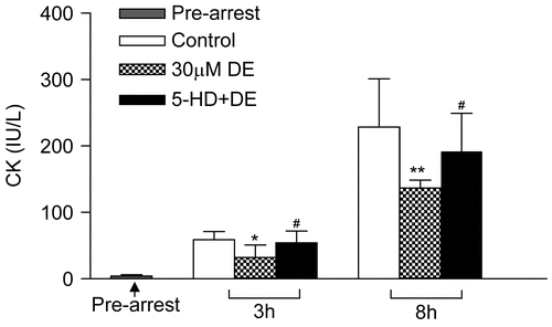
Figure 4.  Release of myocardial creatine kinase (CK) in coronary effluent before arrest and at the 4th min of reperfusion after hypothermic preservation. Data are expressed as mean ± SD, n = 8. *p < 0.05, **p < 0.01 vs. control group; #p < 0.05, ##p < 0.01 vs. 30 μM DE group.

Figure 5. Evaluation of cardiomyocyte apoptosis using terminal dUTP nick-end labeling (TUNEL) after 8 h of hypothermic preservation. Data are expressed as mean ± SD, n = 7–8. *p < 0.05 vs. control group; #p < 0.05 vs. 30 μM DE group.
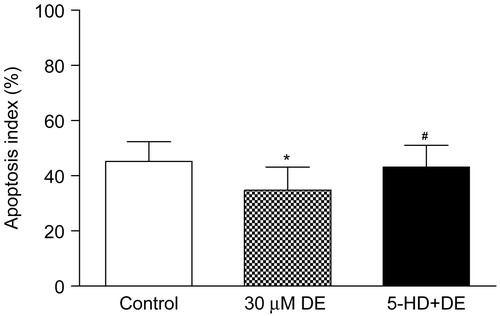
Figure 5.  Evaluation of cardiomyocyte apoptosis using terminal dUTP nick-end labeling (TUNEL) after 8 h of hypothermic preservation. Data are expressed as mean ± SD, n = 7–8. *p < 0.05 vs. control group; #p < 0.05 vs. 30 μM DE group.

Figure 6. Cardiomyocyte apoptosis detected by TUNEL after 8 h hypothermic preservation (×200). Brown staining (TUNEL positive) indicates apoptotic cardiomyocytes (arrow). (A) Normal group: myocardium simply perfused with K–H solution for 30 min; (B) control group: myocardium preserved in Celsior solution for 8 h; (C) DE group: myocardium preserved in Celsior solution supplemented with 30 μM DE for 8 h; (D) 5-HD + DE group: myocardium preserved in Celsior solution supplemented with 30 μM DE and 100 μM 5-HD for 8 h.
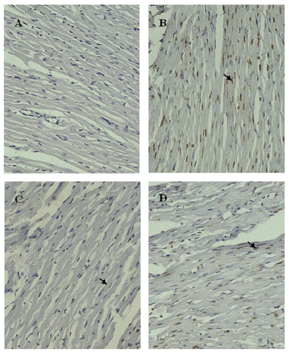
Figure 6.  Cardiomyocyte apoptosis detected by TUNEL after 8 h hypothermic preservation (×200). Brown staining (TUNEL positive) indicates apoptotic cardiomyocytes (arrow). (A) Normal group: myocardium simply perfused with K–H solution for 30 min; (B) control group: myocardium preserved in Celsior solution for 8 h; (C) DE group: myocardium preserved in Celsior solution supplemented with 30 μM DE for 8 h; (D) 5-HD + DE group: myocardium preserved in Celsior solution supplemented with 30 μM DE and 100 μM 5-HD for 8 h.

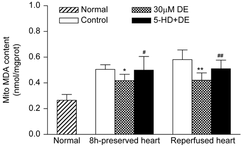
Figure 7. Activities of mitochondrial superoxide dismutase (SOD) in preserved hearts and reperfused hearts. Data are expressed as mean ± SD, n = 8. **p < 0.01 vs. control group; #p < 0.05 vs. 30 μM DE group.Compared with the control, the 30 μM DE addition could significantly inhibit the rise in cardiomyocyte mitochondrial MDA level after 8 h in vitro hypothermic ischemic preservation and 8 h ischemic preservation plus 30 min reperfusion (p < 0.05). The 5-HD + DE group showed no significant difference compared with the control (p > 0.05) ().
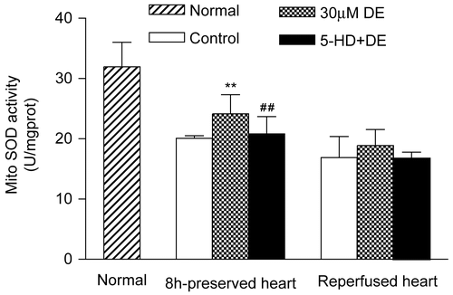
Figure 7.  Activities of mitochondrial superoxide dismutase (SOD) in preserved hearts and reperfused hearts. Data are expressed as mean ± SD, n = 8. **p < 0.01 vs. control group; #p < 0.05 vs. 30 μM DE group.Compared with the control, the 30 μM DE addition could significantly inhibit the rise in cardiomyocyte mitochondrial MDA level after 8 h in vitro hypothermic ischemic preservation and 8 h ischemic preservation plus 30 min reperfusion (p < 0.05). The 5-HD + DE group showed no significant difference compared with the control (p > 0.05) (Figure 8).

Figure 8. Changes of mitochondrial malondialdehyde (MDA) in preserved hearts and reperfused hearts. Data are expressed as mean ± SD, n = 8. *p < 0.05, **p < 0.01 vs. control group; #p < 0.05, ##p < 0.01 vs. 30 μM DE group.

Figure 8.  Changes of mitochondrial malondialdehyde (MDA) in preserved hearts and reperfused hearts. Data are expressed as mean ± SD, n = 8. *p < 0.05, **p < 0.01 vs. control group; #p < 0.05, ##p < 0.01 vs. 30 μM DE group.

Reprints and Corporate Permissions

Please note: Selecting permissions does not provide access to the full text of the article, please see our help page How do I view content?

To request a reprint or corporate permissions for this article, please click on the relevant link below:

Academic Permissions

Please note: Selecting permissions does not provide access to the full text of the article, please see our help page How do I view content?

Obtain permissions instantly via Rightslink by clicking on the button below:

If you are unable to obtain permissions via Rightslink, please complete and submit this Permissions form. For more information, please visit our Permissions help page.