2,100
Views
45
CrossRef citations to date
0
Altmetric
Research Article

Antiproliferative effect of extracts from Aristolochia baetica and Origanum compactum on human breast cancer cell line MCF-7

, , , &
Pages 269-274 | Received 11 Feb 2009, Accepted 07 Jun 2009, Published online: 20 Jan 2010

Figures & data

Table 1. Ethnobotanical data and antiproliferative effect of extracts from the studied plants.

Figure 1. Antiproliferative effect of Aristolochia baetica L extracts on MCF-7 cells. Cells were treated with various concentrations for 24 (—), 48 (-·-), and 72 h (). (A) MCF-7 cells are treated by hexane extract. (B) MCF-7 cells are treated by chloroform extract. (C) MCF-7 cells are treated by ethyl acetate extract. Results are presented as a percentage of negative control (untreated cells) proliferation. Values were expressed as mean ± SD of six experiments (p-value relative to control group: p <0.05).
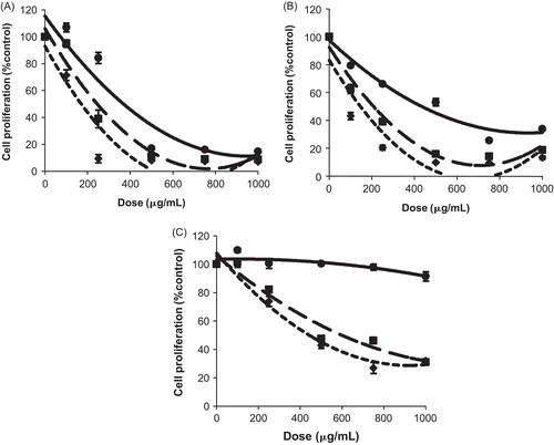
Figure 1.  Antiproliferative effect of Aristolochia baetica L extracts on MCF-7 cells. Cells were treated with various concentrations for 24 (—), 48 (-·-), and 72 h (…). (A) MCF-7 cells are treated by hexane extract. (B) MCF-7 cells are treated by chloroform extract. (C) MCF-7 cells are treated by ethyl acetate extract. Results are presented as a percentage of negative control (untreated cells) proliferation. Values were expressed as mean ± SD of six experiments (p-value relative to control group: p <0.05).
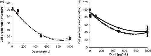
Figure 2. Antiproliferative effect of Origanum compactum Benth. extracts on MCF-7 cells. Cells were treated with various concentrations for 24 h (—), 48 h (-·-), and 72 h (). (A) MCF-7 cells are treated by ethyl acetate extract. (B) MCF-7 cells are treated by methanol extract. Results are presented as percentage of negative control (untreated cells) proliferation. Values were expressed as mean ± SD of six experiments (p-value relative to control group: p <0.05).

Figure 2.  Antiproliferative effect of Origanum compactum Benth. extracts on MCF-7 cells. Cells were treated with various concentrations for 24 h (—), 48 h (-·-), and 72 h (…). (A) MCF-7 cells are treated by ethyl acetate extract. (B) MCF-7 cells are treated by methanol extract. Results are presented as percentage of negative control (untreated cells) proliferation. Values were expressed as mean ± SD of six experiments (p-value relative to control group: p <0.05).

Reprints and Corporate Permissions

Please note: Selecting permissions does not provide access to the full text of the article, please see our help page How do I view content?

To request a reprint or corporate permissions for this article, please click on the relevant link below:

Academic Permissions

Please note: Selecting permissions does not provide access to the full text of the article, please see our help page How do I view content?

Obtain permissions instantly via Rightslink by clicking on the button below:

If you are unable to obtain permissions via Rightslink, please complete and submit this Permissions form. For more information, please visit our Permissions help page.