944
Views
15
CrossRef citations to date
0
Altmetric
Research Article

Immunomodulatory effects of damnacanthal isolated from roots of Morinda elliptica

, , , , &
Pages 446-452 | Received 04 Jul 2008, Accepted 04 Feb 2009, Published online: 24 Mar 2010

Figures & data

Figure 1. Mitogenic activity of damnacanthal on mouse thymocytes was determined by using MTT assay. Mouse thymocytes were isolated and incubated with increasing concentrations (0.468 μg/mL–30 μg/mL) of damnacanthal or Con A (2.5 μg/mL, as positive control) in culture medium for 24, 48, and 72h. Results were expressed as mean percentage ratio of MTT absorbance in damnacanthal-treated and control well ± standard error of three independent experiments with three wells each.
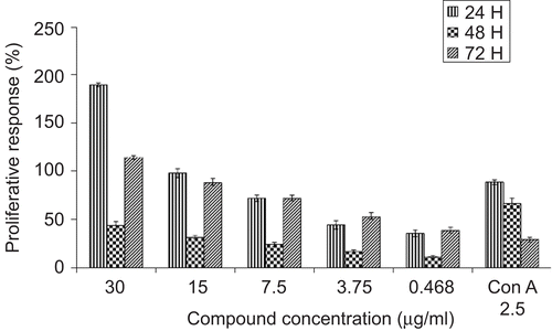
Figure 1.  Mitogenic activity of damnacanthal on mouse thymocytes was determined by using MTT assay. Mouse thymocytes were isolated and incubated with increasing concentrations (0.468 μg/mL–30 μg/mL) of damnacanthal or Con A (2.5 μg/mL, as positive control) in culture medium for 24, 48, and 72h. Results were expressed as mean percentage ratio of MTT absorbance in damnacanthal-treated and control well ± standard error of three independent experiments with three wells each.
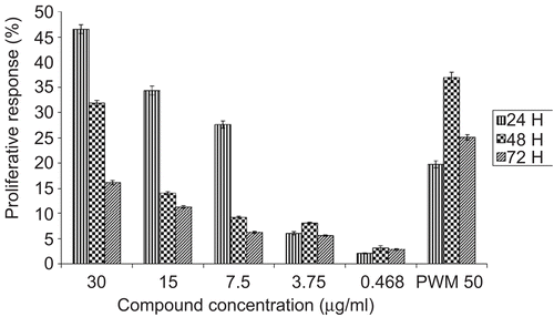
Figure 2. Mitogenic activity of damnacanthal on PBMC was tested by using MTT assay. PBMC were isolated and incubated with increasing concentrations (0.468 μg/mL–30 μg/mL) of damnacanthal or PWM (50 μg/mL, as positive control) in culture medium for 24, 48, and 72h. Results were expressed as mean percentage ratio of MTT absorbance in damnacanthal-treated and control well ± standard error of three independent experiments with three wells each.

Figure 2.  Mitogenic activity of damnacanthal on PBMC was tested by using MTT assay. PBMC were isolated and incubated with increasing concentrations (0.468 μg/mL–30 μg/mL) of damnacanthal or PWM (50 μg/mL, as positive control) in culture medium for 24, 48, and 72h. Results were expressed as mean percentage ratio of MTT absorbance in damnacanthal-treated and control well ± standard error of three independent experiments with three wells each.

Table 1. Flow cytometry analysis of cell cycle distribution on PBMC based on proliferation effect of damnacanthal and PWM. The values were the means ± SE of three experiments.

Figure 3. The production of human interleukin-2 in culture supernatants upon stimulation of PBMC by damnacanthal, and PWM. PBMC were isolated and incubated at 24, 48, and 72 h with active concentrations of damnacanthal at 30 μg/mL, while PWM at 50 μg/mL and interleukin-2 induction was specifically determined by ELISA.
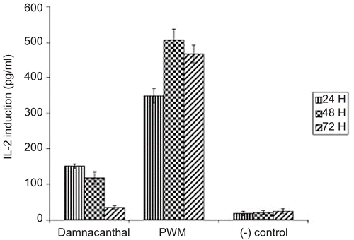
Figure 3.  The production of human interleukin-2 in culture supernatants upon stimulation of PBMC by damnacanthal, and PWM. PBMC were isolated and incubated at 24, 48, and 72 h with active concentrations of damnacanthal at 30 μg/mL, while PWM at 50 μg/mL and interleukin-2 induction was specifically determined by ELISA.
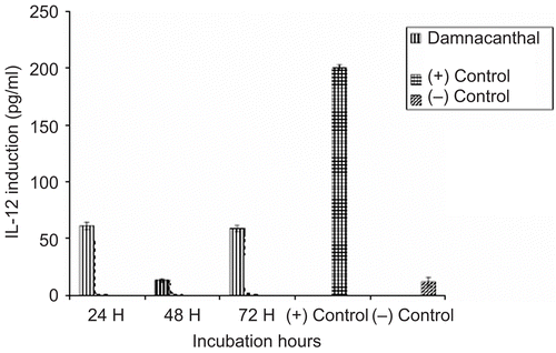
Figure 4. The production of human interleukin-12 in culture supernatants upon stimulation of PBMC by damnacanthal. PBMC were isolated and incubated at 24, 48, and 72 h with active concentrations (damnacanthal at 30 μg/mL) and interleukin-2 induction was specifically determined by ELISA.

Figure 4.  The production of human interleukin-12 in culture supernatants upon stimulation of PBMC by damnacanthal. PBMC were isolated and incubated at 24, 48, and 72 h with active concentrations (damnacanthal at 30 μg/mL) and interleukin-2 induction was specifically determined by ELISA.

Reprints and Corporate Permissions

Please note: Selecting permissions does not provide access to the full text of the article, please see our help page How do I view content?

To request a reprint or corporate permissions for this article, please click on the relevant link below:

Academic Permissions

Please note: Selecting permissions does not provide access to the full text of the article, please see our help page How do I view content?

Obtain permissions instantly via Rightslink by clicking on the button below:

If you are unable to obtain permissions via Rightslink, please complete and submit this Permissions form. For more information, please visit our Permissions help page.