Figures & data
Table 1. Evaluation of erythema and exudate.
Figure 1. The average size of lesions infected with C. albicans (A), C. neoformans (B), M. canis (C), and S. schenckii (D) and treated with four extracts, isolated compound, and amphotericin B (positive control).
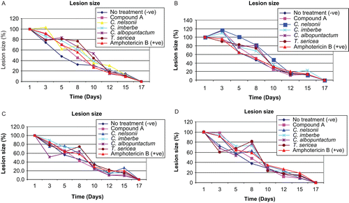
Figure 2. The influence of the crude extracts, isolated compound, and amphotericin B (positive control) on wound erythema in rats infected with C. albicans (A), C. neoformans (B), M. canis (C), and S. schenckii (D).
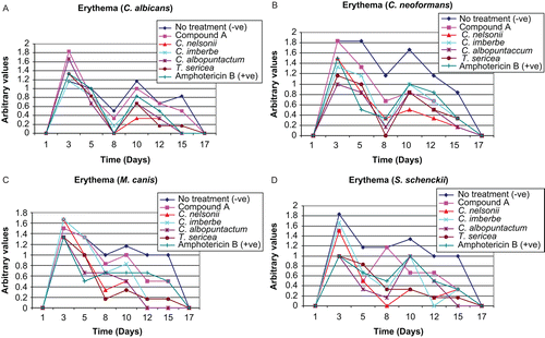
Figure 3. The influence of the crude extracts, isolated compound, and amphotericin B (positive control) on exudate formed in rats infected with C. albicans (A), C. neoformans (B), M. canis (C), and S. schenckii (D).
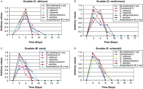
Table 2. Quantitative histopathological findings of wounds of rats infected with C. albicans after topical application of different creams.
Table 3. Quantitative histopathological findings of wounds of rats infected with C. neoformans after topical application of different creams.
Table 4. Quantitative histopathological findings of wounds of rats infected with M. canis after topical application of different creams.
Table 5. Quantitative histopathological findings of wounds of rats infected with S. schenckii after topical application of different creams.
Table 6. Minimum inhibitory concentration (MIC) values of selected extracts after 24 h incubation at 37°C.
Table 7. LC50 values from MTT cytotoxicity assay of the optimal extracts.