Figures & data
Figure 1. Ball and stick model with labels of optimized structure of the β-sitosterol molecule. Red: oxygen; dark gray: carbon; light gray: hydrogen (PM3 result).
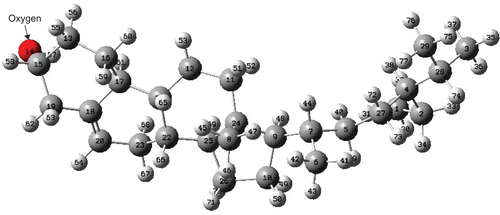
Figure 2. Bond lengths between carbon atoms in β-sitosterol molecule; hydrogen atoms are not displayed (PM3 result).
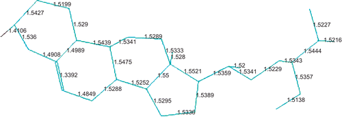
Table 1. Energy contributions (in kcal/mol) after molecular mechanics (MM) method with MM+ force field.
Table 2. Calculated energies (in kcal/mol) after PM3 method.
Figure 3. Calculated infrared spectrum of β-sitosterol molecule (PM3 result).
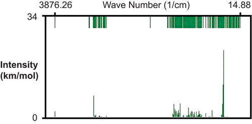
Figure 4. Directions of vibrations in β-sitosterol molecule (PM3 result).
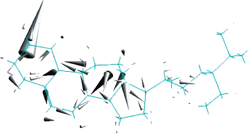
Table 3. The first 16 relatively larger infrared intensities (in km/mol) and the corresponding harmonic frequencies (in cm−1) after PM3 method.
Table 4. Some of the calculated quantities after DFT/B3LYP/6-31G* method.
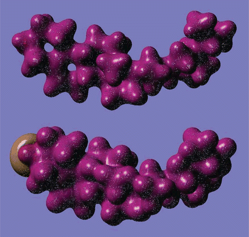
Figure 5. 3D images of highest occupied molecular orbital (HOMO, upper) and lowest unoccupied molecular orbital (LUMO, lower) of β-sitosterol molecule (DFT/6-31G* results).
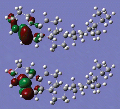
Figure 6. Excess (Mulliken) charge on atoms of β-sitosterol molecule (DFT/6-31G* result).
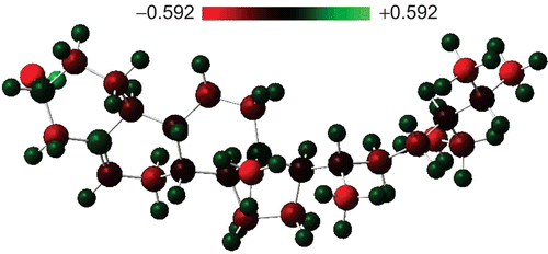
Figure 7. 3D images of charge density (upper) and electrostatic potential (lower) of β-sitosterol molecule (DFT/6-31G* results).