976
Views
4
CrossRef citations to date
0
Altmetric
Research Article

Improving the absorption of earthworm fibrinolytic enzymes with mucosal enhancers

, &
Pages 816-821 | Received 23 Apr 2009, Accepted 08 Jun 2009, Published online: 03 Jun 2010

Figures & data

Figure 1. Cytotoxic effects of the six enhancers on Caco-2 cell monolayers. Cytotoxicity was measured by the MTT assay following 2, 6, and 24 h incubation at 37°C, 5% CO2 and 95% relative humidity. For the MTT assay, PBS and DMSO were used as positive (100% cell viability) and negative (0% cell viability) controls, respectively. All measurements were expressed as mean ± SD.
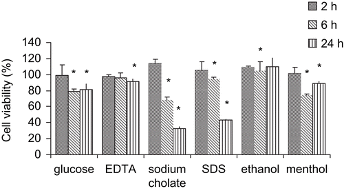
Figure 1.  Cytotoxic effects of the six enhancers on Caco-2 cell monolayers. Cytotoxicity was measured by the MTT assay following 2, 6, and 24 h incubation at 37°C, 5% CO2 and 95% relative humidity. For the MTT assay, PBS and DMSO were used as positive (100% cell viability) and negative (0% cell viability) controls, respectively. All measurements were expressed as mean ± SD.

Figure 2. Cumulative permeation of EFEs across the Caco-2 cell monolayers. Six different enhancers were added to the apical compartment of the insert individually to increase the EFEs permeation.
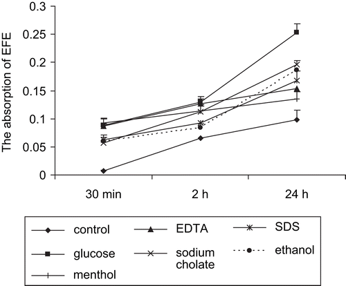
Figure 2.  Cumulative permeation of EFEs across the Caco-2 cell monolayers. Six different enhancers were added to the apical compartment of the insert individually to increase the EFEs permeation.
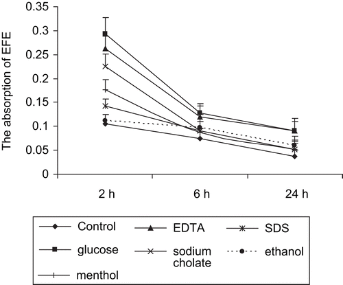
Figure 3. FITC-labeled EFEs and different enhancers were simultaneously intragastrically administrated to mice. After 2, 6, and 24 h, serum samples were obtained and counted by Bio-Tek fluorescence spectrophotometer.

Figure 3.  FITC-labeled EFEs and different enhancers were simultaneously intragastrically administrated to mice. After 2, 6, and 24 h, serum samples were obtained and counted by Bio-Tek fluorescence spectrophotometer.

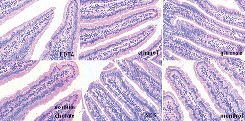
Figure 4. Light micrographs of microvilli from duodenum epithelia tissues at 24 h after oral administration of the enhancers in healthy mice (×400).

Figure 4.  Light micrographs of microvilli from duodenum epithelia tissues at 24 h after oral administration of the enhancers in healthy mice (×400).

Reprints and Corporate Permissions

Please note: Selecting permissions does not provide access to the full text of the article, please see our help page How do I view content?

To request a reprint or corporate permissions for this article, please click on the relevant link below:

Academic Permissions

Please note: Selecting permissions does not provide access to the full text of the article, please see our help page How do I view content?

Obtain permissions instantly via Rightslink by clicking on the button below:

If you are unable to obtain permissions via Rightslink, please complete and submit this Permissions form. For more information, please visit our Permissions help page.