1,170
Views
14
CrossRef citations to date
0
Altmetric
Research Article

Protective effects of Pergularia daemia roots against paracetamol and carbon tetrachloride-induced hepatotoxicity in rats

&
Pages 1265-1272 | Received 28 Sep 2009, Accepted 24 Feb 2010, Published online: 15 Sep 2010

Figures & data

Table 1. Phytochemical screening of Pergularia daemia root extracts (PdAE and PdEE).

Table 2. Effect of PdAE and PdEE on serum enzyme and biochemical parameters in paracetamol-induced hepatotoxicity in rats.

Table 3. Effect of PdAE and PdEE on liver weight, glutathione (GSH) and lipid peroxidation (LPO) in paracetamol-induced hepatotoxicity in rats.

Table 4. Effect of PdAE and PdEE on serum enzyme and biochemical parameters in CCL4-induced hepatotoxicity in rats.

Table 5. Effect of PdAE and PdEE on liver weight, glutathione (GSH) and lipid peroxidation (LPO) in CCL4-induced hepatotoxicity in rats.

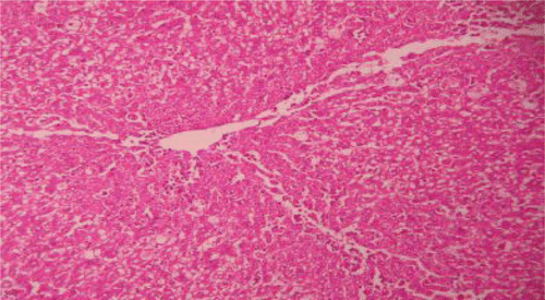
Figure 1. Liver section of control rats showing normal hepatic cells.

Figure 1.  Liver section of control rats showing normal hepatic cells.

Figure 2. Liver section of paracetamol rats shows necrosis, ballooning degeneration and massive infiltration of lymphocytes.
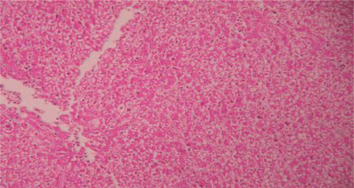
Figure 2.  Liver section of paracetamol rats shows necrosis, ballooning degeneration and massive infiltration of lymphocytes.

Figure 3. Liver section of silymarin and paracetamol rats shows moderate necrosis, moderate ballooning degeneration and little infiltration of lymphocytesries.
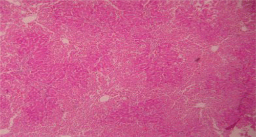
Figure 3.  Liver section of silymarin and paracetamol rats shows moderate necrosis, moderate ballooning degeneration and little infiltration of lymphocytesries.

Figure 4. Liver section of PdAE (200 mg/kg) and paracetamol showing less fatty changes, mild necrosis, degeneration and little infiltration of lymphocytes.
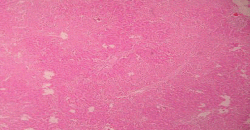
Figure 4.  Liver section of PdAE (200 mg/kg) and paracetamol showing less fatty changes, mild necrosis, degeneration and little infiltration of lymphocytes.

Figure 5. Liver section of PdEE (200 mg/kg) and paracetamol showing moderate fatty changes, necrosis, ballooning degeneration and no infiltration of lymphocytes.
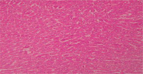
Figure 5.  Liver section of PdEE (200 mg/kg) and paracetamol showing moderate fatty changes, necrosis, ballooning degeneration and no infiltration of lymphocytes.

Figure 6. Liver section of CCl4 rats showing fatty changes, necrosis and filtration of lymphocytes.

Figure 6.  Liver section of CCl4 rats showing fatty changes, necrosis and filtration of lymphocytes.

Figure 7. Liver section of silymarin and CCl4shows mild fatty changes, necrosis, mild ballooning degeneration and mild infiltration of lymphocytes.
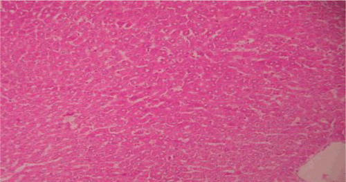
Figure 7.  Liver section of silymarin and CCl4shows mild fatty changes, necrosis, mild ballooning degeneration and mild infiltration of lymphocytes.
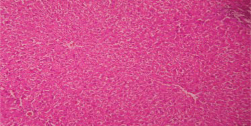
Figure 8. Liver section of PdAE (200 mg/kg) and CCl4 shows mild fatty changes, necrosis, mild ballooning with degeneration and mild infiltration of lymphocytes.
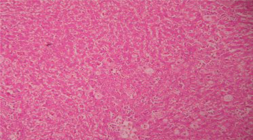
Figure 8.  Liver section of PdAE (200 mg/kg) and CCl4 shows mild fatty changes, necrosis, mild ballooning with degeneration and mild infiltration of lymphocytes.
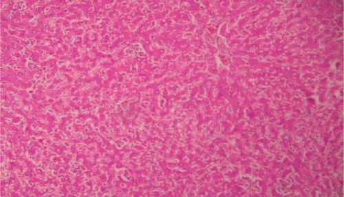
Figure 9. Liver section of PdEE (200 mg/kg) and CCl4 shows fatty changes necrosis, ballooning degeneration and few infiltrations of lymphocytes.

Figure 9.  Liver section of PdEE (200 mg/kg) and CCl4 shows fatty changes necrosis, ballooning degeneration and few infiltrations of lymphocytes.

Reprints and Corporate Permissions

Please note: Selecting permissions does not provide access to the full text of the article, please see our help page How do I view content?

To request a reprint or corporate permissions for this article, please click on the relevant link below:

Academic Permissions

Please note: Selecting permissions does not provide access to the full text of the article, please see our help page How do I view content?

Obtain permissions instantly via Rightslink by clicking on the button below:

If you are unable to obtain permissions via Rightslink, please complete and submit this Permissions form. For more information, please visit our Permissions help page.