Figures & data
Figure 1. Chemical structures of compounds isolated from M. exotica. They were confirmed by 1H NMR, 13C NMR, MS and IR spectra and comparing with reference data from available literature (CitationIto et al., 1987; CitationBulut et al., 1996; CitationWu et al., 1989).
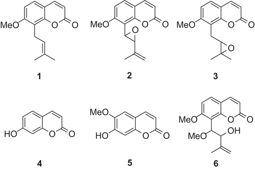
Figure 2. Extraction by different solvents for anti-nociceptive activities. Results are expressed as mean ± SD (n = 6). *P < 0.05 and **P < 0.01 compared with control.
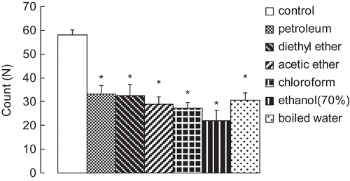
Figure 3. (A) Effect of the ethanol extracts on acetic acid-induced writhing response in mice. (B) Effect of the isolated compounds on acetic acid-induced writhing response in mice. Results are expressed as mean ± SD (n = 6), *P < 0.05 and **P < 0.01 compared with control.
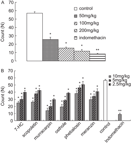
Figure 4. Effect of the ethanol extracts on hot-plate test in mice. Results are expressed as mean ± SD (n = 6), *P < 0.05 and **P < 0.01 compared with control.
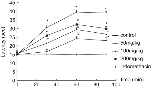
Table 1. Effect of the isolated compounds on hot-plate test in mice.
Figure 5. (A) Inhibitory action of the ethanol extracts on carrageenan- induced paw edema in rats. (B) Inhibitory action of the isolated compounds on carrageenan-induced paw edema in rat. Results are expressed as mean ± SD, (n = 6), *P < 0.05 and **P < 0.01 compared with control.
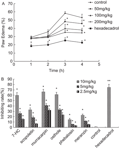
Figure 6. (A) Effect of ethanol extract on xylene-induced ear edema in mice. (B) Effect of the isolated compounds on xylene-induced ear edema in mice. Results are expressed as mean ± SD, (n = 6),*P < 0.05 and **P < 0.01 compared with control.
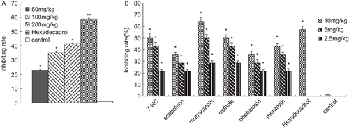
Figure 7. The gross observation and the histomorphology of femoral condyle in knee joint. (A) and (D) are the control groups; (B) and (E) are the model groups, which demonstrated the severe ulcer (see the arrow) in femoral condyle. In this ulcer area, hyperplasia was seen in the synovium and the other cell layers were unclear. (C) and (F) are the ethanol extract (200 mg/kg) groups. There were no obvious differences when compared with the control groups, indicating regular surface and good arrangement of all cell layers. However, there was a little congestion in (C) and hypertrophy and a little shorter height in cartilage in (F).
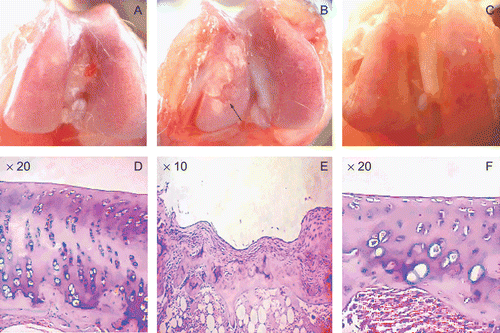
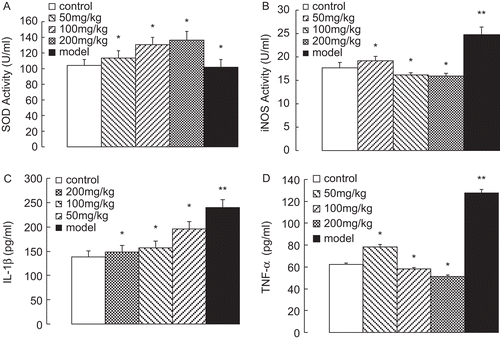
Figure 8. (A) SOD activity, (B) iNOS activity, (C) IL-1β content and (D) TNF-α content in rat serum after an eight week establishment of OA models. Results are expressed as mean ± SD (n = 6), *P < 0.05 and **P < 0.01 compared with control.